Ever wondered what the world looks like beyond what the naked eye can see? The 2025 Nikon Small World Photomicrography Competition just dropped its winners, and trust us—these images will blow your mind. For over 50 years, Nikon Small World has been the go-to stage for showcasing the mind-bending beauty and complexity of life under the light microscope.
This year’s winning shots are pure magic—tiny worlds exploding with color, structure, and hidden stories. From the delicate wings of a butterfly to the glowing neurons of a zebrafish embryo, each photo reveals the insane artistry built into nature itself. What’s wild is that many of these jaw-dropping visuals weren’t made by pros with fancy studios—they came from scientists, students, and everyday enthusiasts who simply share a passion for seeing life differently.
The Small World in Motion category also turned heads with its hypnotic time-lapse videos that show cells dividing, organisms dancing, and microscopic landscapes pulsing with energy. Every frame is a fusion of science and art, reminding us that the tiniest things can hold the biggest wonders.
The Nikon Small World competition isn’t just about pretty pictures—it’s about curiosity, creativity, and the drive to explore what lies beneath the surface. It celebrates a global community of visual storytellers who turn data into emotion and science into beauty. These 20 winners prove that when you mix passion with precision, the result is nothing short of mind-blowing.
You can find more info about Nikon Small World:
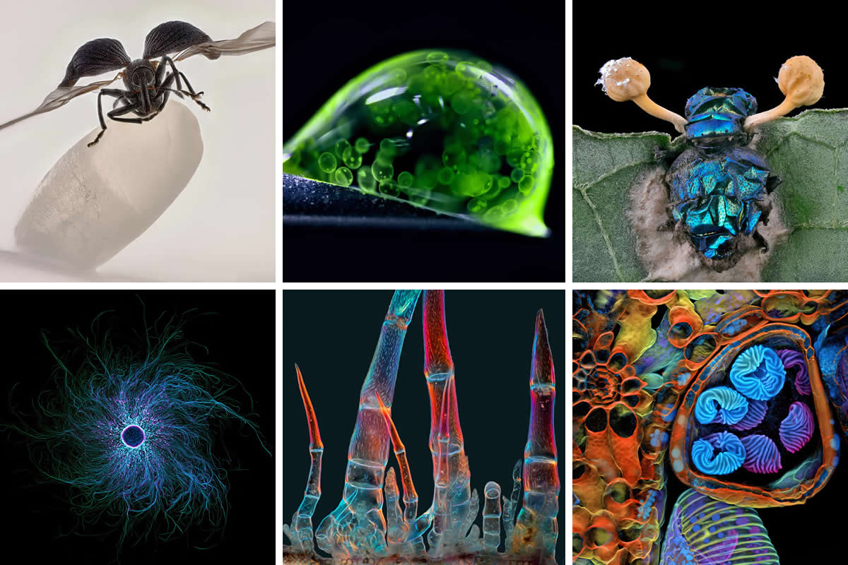
#1. 1st Place: "Rice weevil (Sitophilus oryzae) on a grain of rice" by Zhang You
- Location: Kunming, Yunnan, China
- Technique: Image Stacking
- Magnification: 5X (Objective Lens Magnification)
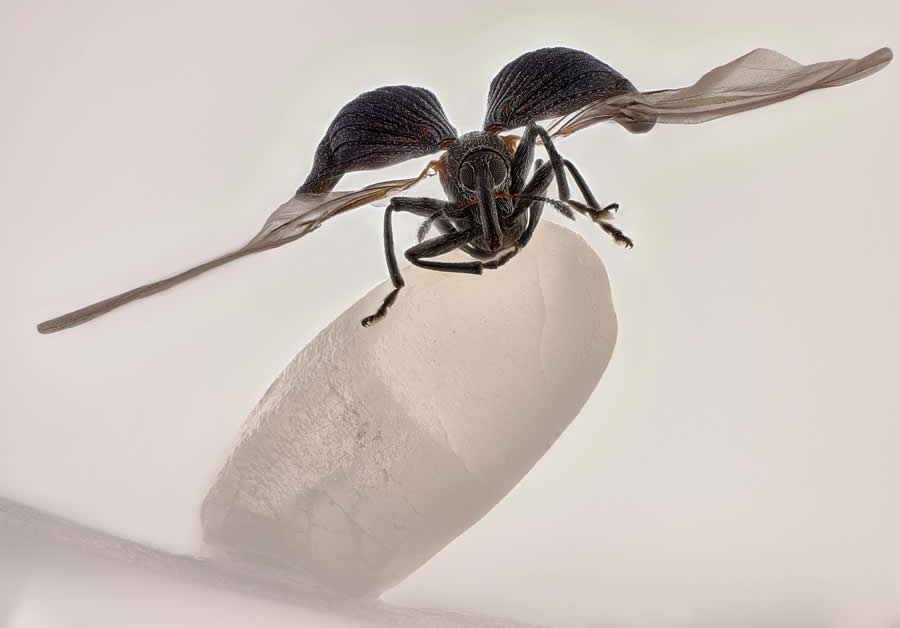
#2. 2nd Place: "Colonial algae (Volvox) spheres in a drop of water" by Dr. Jan Rosenboom
- Location: Rostock, Mecklenburg Vorpommern, Germany
- Technique: Reflected Light
- Magnification: 5X (Objective Lens Magnification)
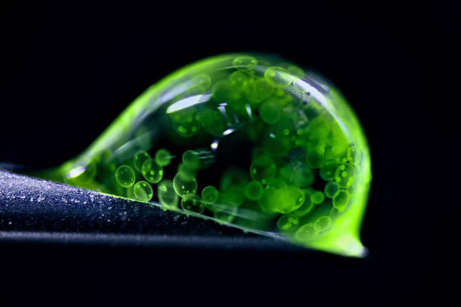
#3. 3rd Place: "Pollen in a garden spider web" by John-Oliver Dum
- Affiliation: Medienbunker Produktion, Bendorf, Rheinland Pfalz, Germany
- Technique: Image Stacking
- Magnification: 20X (Objective Lens Magnification)
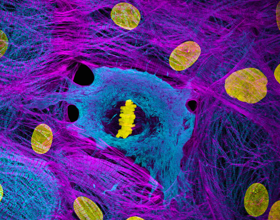
#4. 4th Place: "Heart muscle cells with chromosomes condensed following cell division" by Dr. James Hayes
- Affiliation: Vanderbilt University, Department of Cell and Developmental Biology, Nashville, Tennessee, USA
- Technique: Confocal
- Magnification: 100X (Objective Lens Magnification)
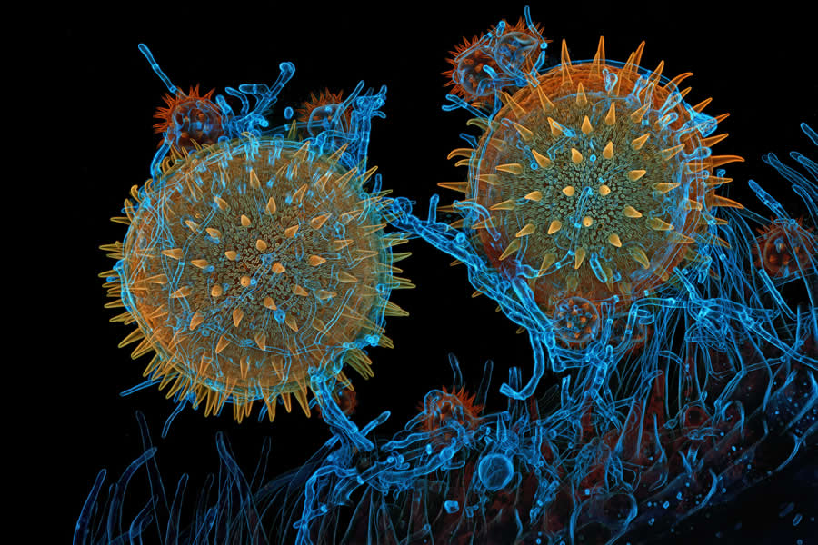
#5. 5th Place: "Spores (blue/purple structures) of a small tropical fern (Ceratopteris richardii)" by Dr. Igor Robert Siwanowicz
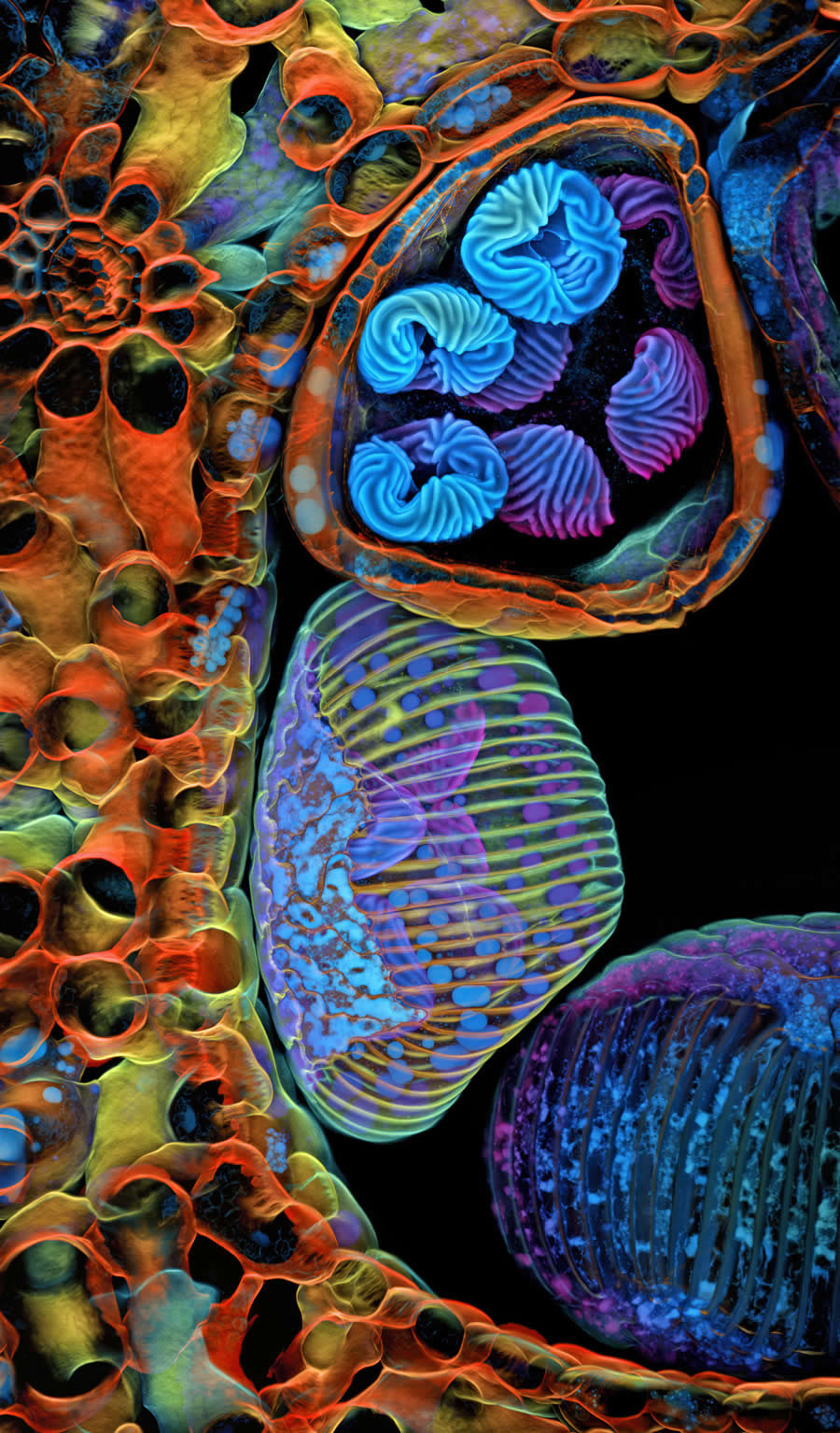
- Affiliation: Howard Hughes Medical Institute (HHMI), Janelia Research Campus, Ashburn, Virginia, USA
- Technique: Confocal
- Magnification: 25X (Objective Lens Magnification)
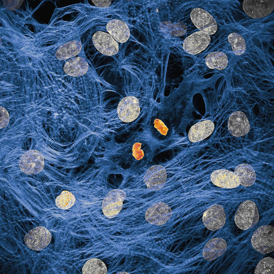
#6. 6th Place: "Rat liver cells" by Dr. Francisco Lázaro-Diéguez
- Affiliation: Albert Einstein College of Medicine, Bronx, New York, USA
- Technique: Confocal
- Magnification: 63X (Objective Lens Magnification)
#7. 7th Place: "iPSC-derived sensory neurons labelled to show tubulin and actin" by Stella Whittaker
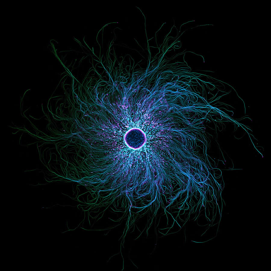
- Affiliation: National Institutes of Health (NIH), National Institute of Neurological Disorders and Stroke, Bethesda, Maryland, USA
- Technique: Confocal, Fluorescence, Image Stacking
- Magnification: 10X (Objective Lens Magnification)
#8. 8th Place: "Mallow pollen germinating on stigma while being parasitized by a filamentous fungus" by Dr. Igor Robert Siwanowicz
- Affiliation: Howard Hughes Medical Institute (HHMI), Janelia Research Campus, Ashburn, Virginia, USA
- Technique: Confocal
- Magnification: 40X (Objective Lens Magnification)
#9. 9th Place: "A fungus (Talaromyces purpureogenus) known for its red, diffused pigment" by Wim van Egmond
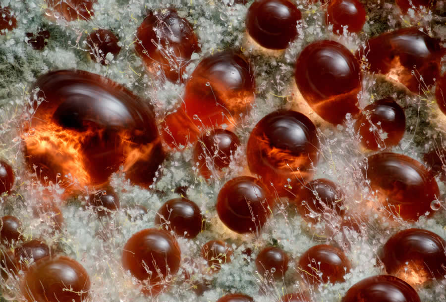
- Affiliation: Micropolitan Museum, Berkel en Rodenrijs, Zuid-Holland, The Netherlands
- Technique: Image Stacking
- Magnification: 10X (Objective Lens Magnification)
#10. 10th Place: "Heart muscle cells (iPSC-derived) showing condensed chromosomes in metaphase" by Dr. Dylan T. Burnette & Dr. James Hayes
- Affiliation: Vanderbilt University, Department of Cell and Developmental Biology, Nashville, Tennessee, USA
- Technique: Structured Illumination Microscopy (SIM)
- Magnification: 60X (Objective Lens Magnification)
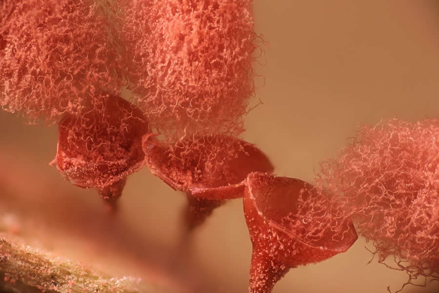
#11. 11th Place: "Sunflower trichomes (hair-like plant outgrowths)" by Marek Miś
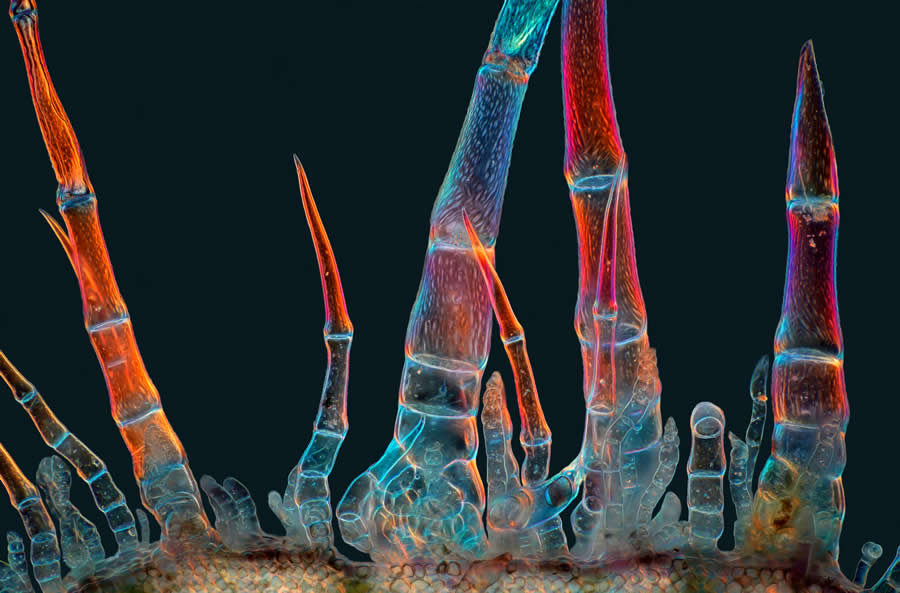
- Affiliation: Marek Mis Photography, Suwalki, Podlaskie, Poland
- Technique: Polarized Light
- Magnification: 10X (Objective Lens Magnification)
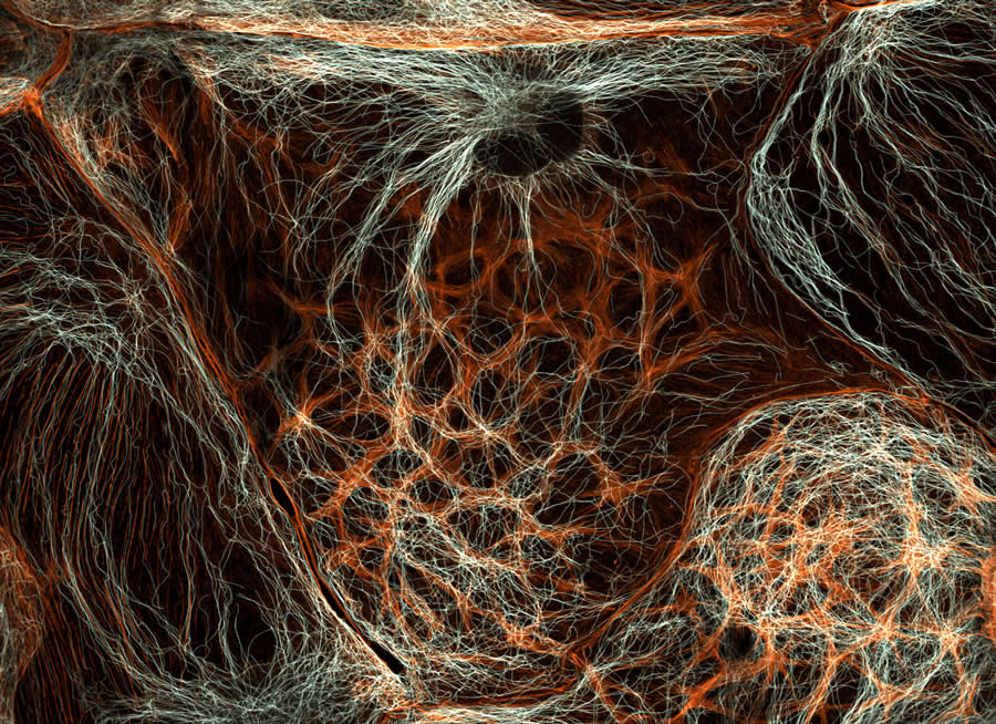
#12. 12th Place: "The actin cytoskeleton (cyan) and endoplasmic reticulum (red) of a mouse brain cancer cell" by Halli Lindamood & Dr. Eric Vitriol
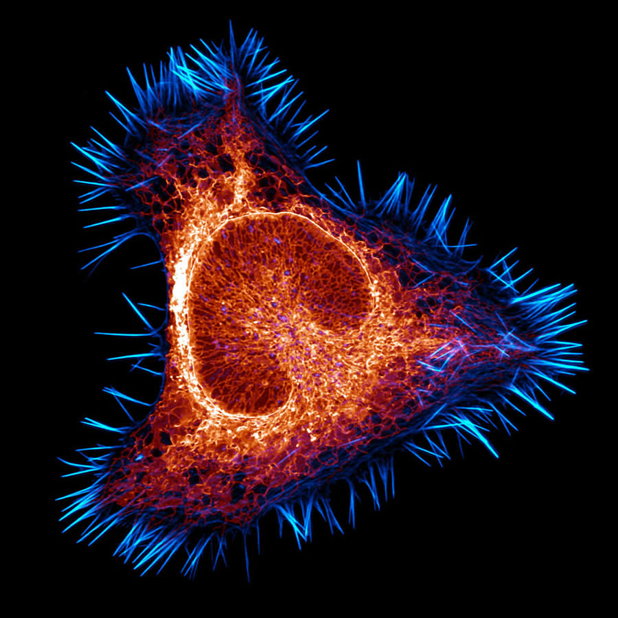
- Affiliation: Augusta University, Department of Neuroscience and Regenerative Medicine, Augusta, Georgia, USA
- Technique: Confocal, Deconvolution
- Magnification: 100X (Objective Lens Magnification)
#13. 13th Place: "Slime mold (Arcyria major) releasing spores" by Henri Koskinen
- Affiliation: University of Helsinki, Helsinki, Uudenmaan lääni, Finland
- Technique: Image Stacking, Reflected Light
- Magnification: 10X (Objective Lens Magnification)
#14. 14th Place: "Quartz with biotic goethite filaments" by Manfred Heising
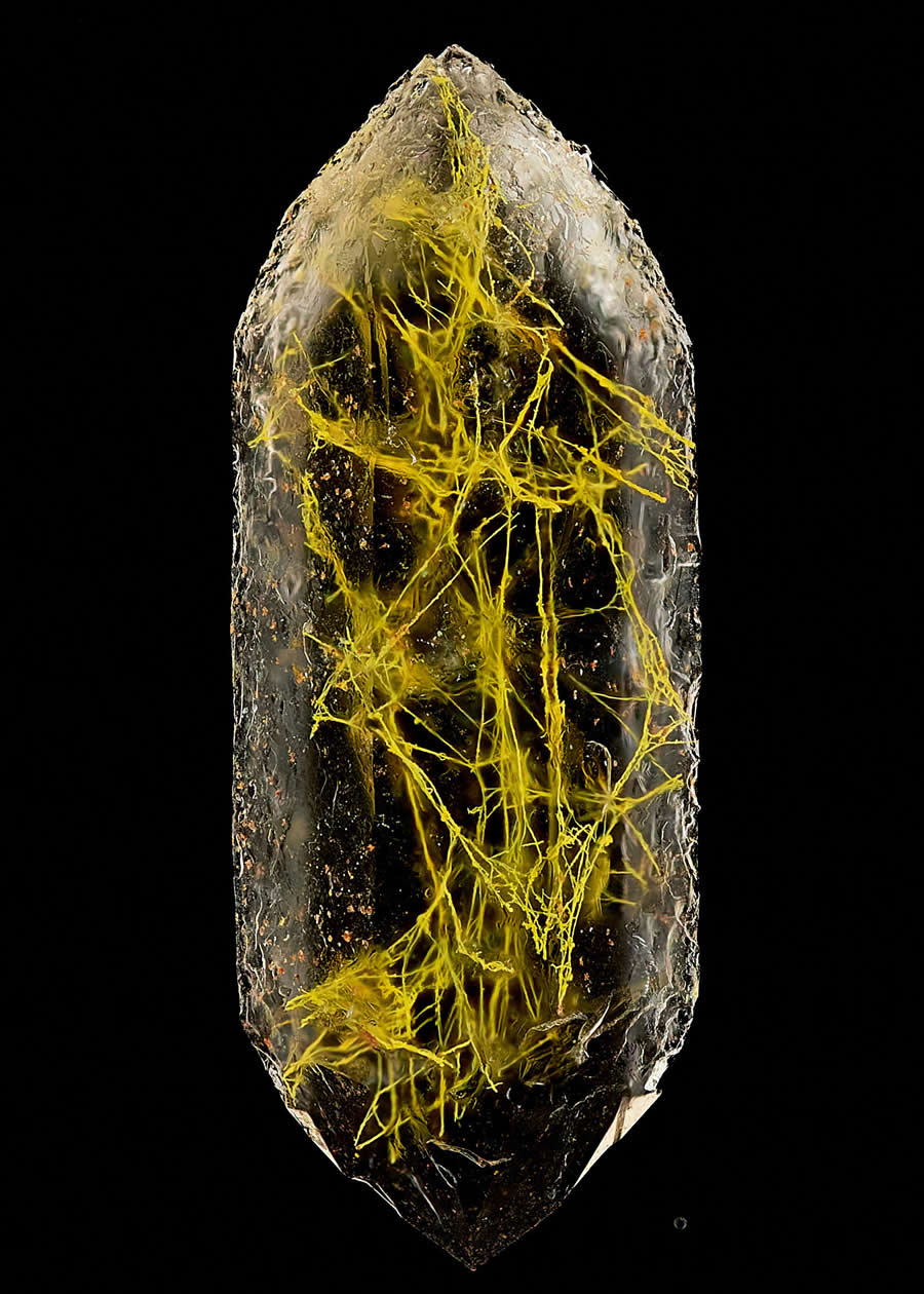
- Affiliation: LWL Museum of Natural History Münster, Münster, Northrhine-Westphalia, Germany
- Technique: Image Stacking
- Magnification: 5X (Objective Lens Magnification)
#15. 15th Place: "Geometer moth (Geometridae) laying eggs" by Zhang You
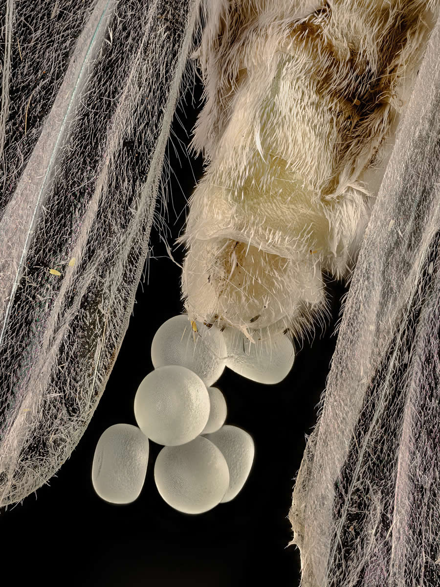
- Location: Kunming, Yunnan, China
- Technique: Image Stacking
- Magnification: 5X (Objective Lens Magnification)
#16. 16th Place: "Spore sacs (sporangia) of a fern" by Rogelio Moreno
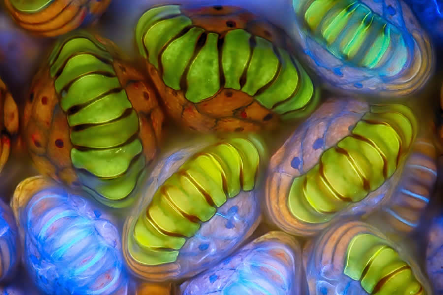
- Location: Panama, Panama
- Technique: Fluorescence, Image Stacking
- Magnification: 40X (Objective Lens Magnification)
#17. 17th Place: "Water fleas (Daphnia) and algae" by Hong Guo
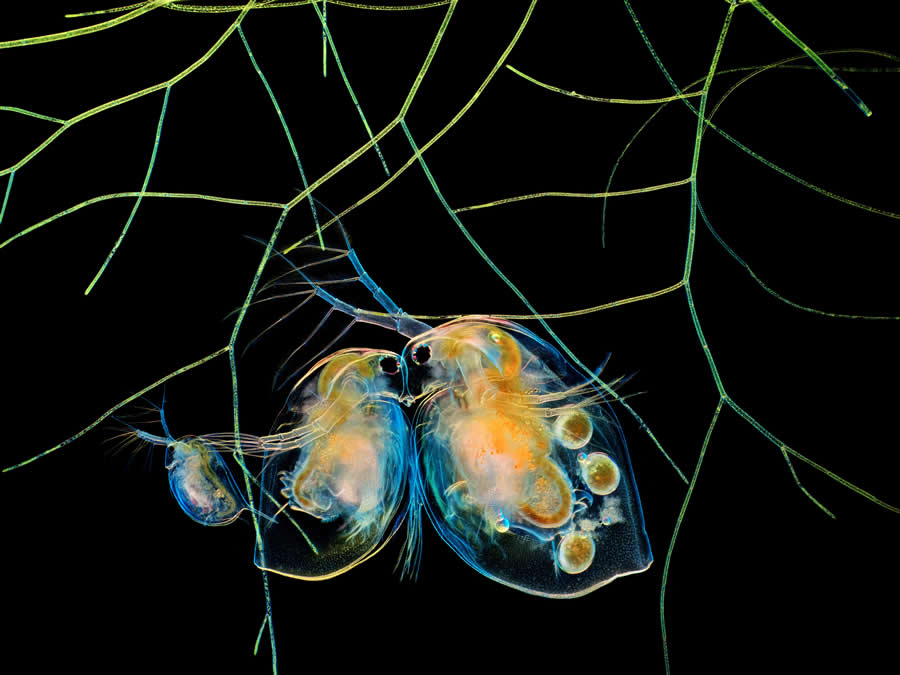
- Location: Chengdu, Si Chuan, China
- Technique: Image Stacking
- Magnification: 5X (Objective Lens Magnification)
#18. 18th Place: "Fluorescently marked mouse colon" by Marius Mählen, Koen Oost, Prisca Liberali, Laurent Gelman
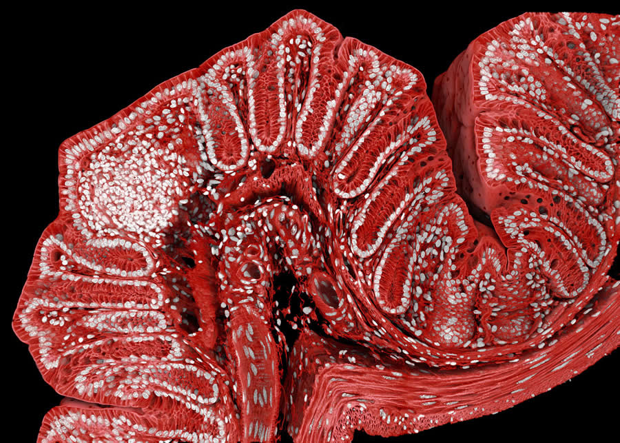
- Affiliation: Friedrich Miescher Institute for Biomedical Research, Basel, Switzerland
- Technique: Confocal
- Magnification: 20X (Objective Lens Magnification)
#19. 19th Place: "Parasitic fungus (Cordycipitaceae) on a fly (Calliphoridae)" by Eduardo Agustin Carrasco
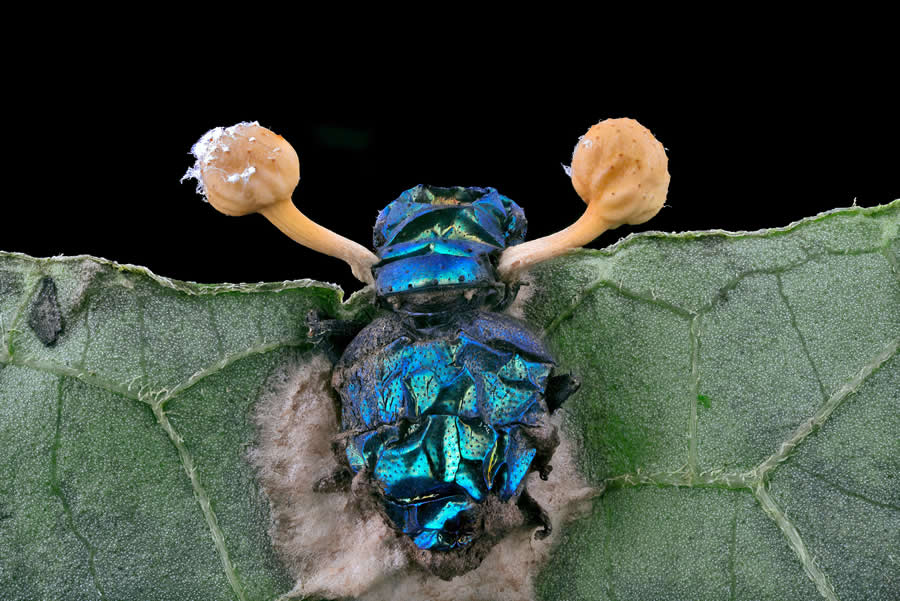
- Location: Cuenca, Azuay, Ecuador
- Technique: Image Stacking
- Magnification: 2X (Objective Lens Magnification)
#20. 20th Place: "Marine copepod" by Dr. Zachary Sanchez
- Affiliation: Vanderbilt University, Department of Cell and Developmental Biology, Nashville, Tennessee, USA
- Technique: Confocal
- Magnification: 60X (Objective Lens Magnification)


