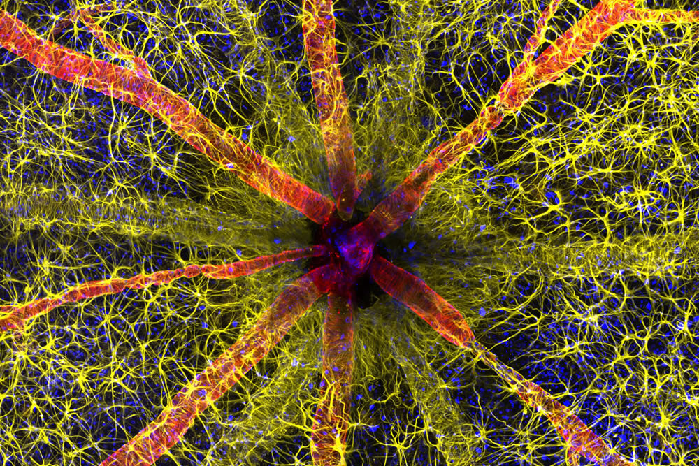
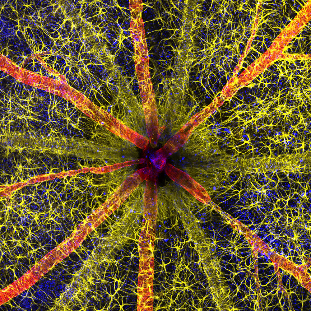
Photomicrography Competition has unveiled its remarkable winners, with Hassanain Qambari clinching the top spot for his stunning portrayal of a rodent optic nerve head.
Initiated in 1975, the Nikon Small World Competition was conceived to honor the dedication of individuals in the realm of light microscope photography. Over the years, it has transformed into a premier platform, showcasing the extraordinary work of photomicrographers from diverse scientific fields. This year, the competition witnessed an impressive participation, receiving close to 1,900 captivating photo entries from 72 countries, reinforcing its status as a global celebration of microscopic artistry.
Scroll down and inspire yourself. You can check their website for more information.
You can find more info about Nikon’s Small World:
#1. Rodent optic nerve head showing astrocytes (yellow), contractile proteins (red) and retinal vasculature (green) by Hassanain Qambari & Jayden Dickson
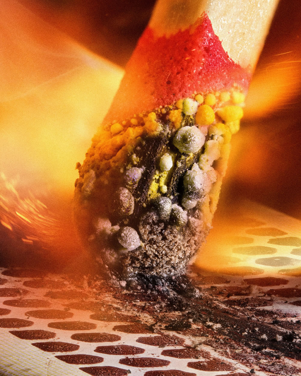
#2. Matchstick igniting by the friction surface of the box by Ole Bielfeldt
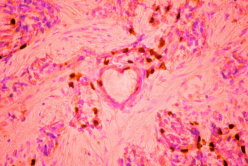
#3. Breast cancer cells by Malgorzata Lisowska
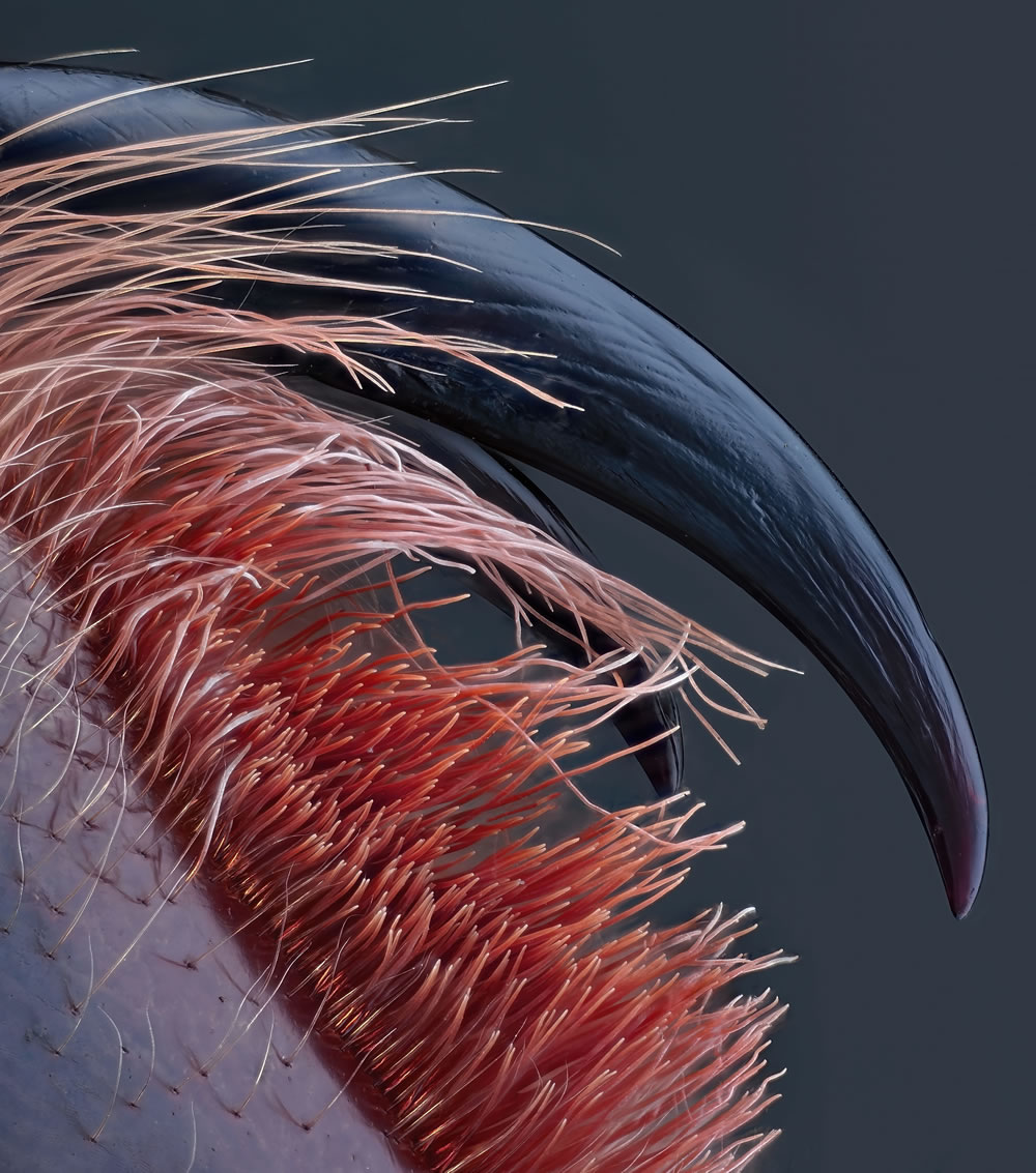
#4. Venomous fangs of a small tarantula by John-Oliver Dum
#5. Auto-fluorescing defensive hairs covering the leaf surface of Eleagnus angustifolia exposed to UV light by Dr. David Maitland
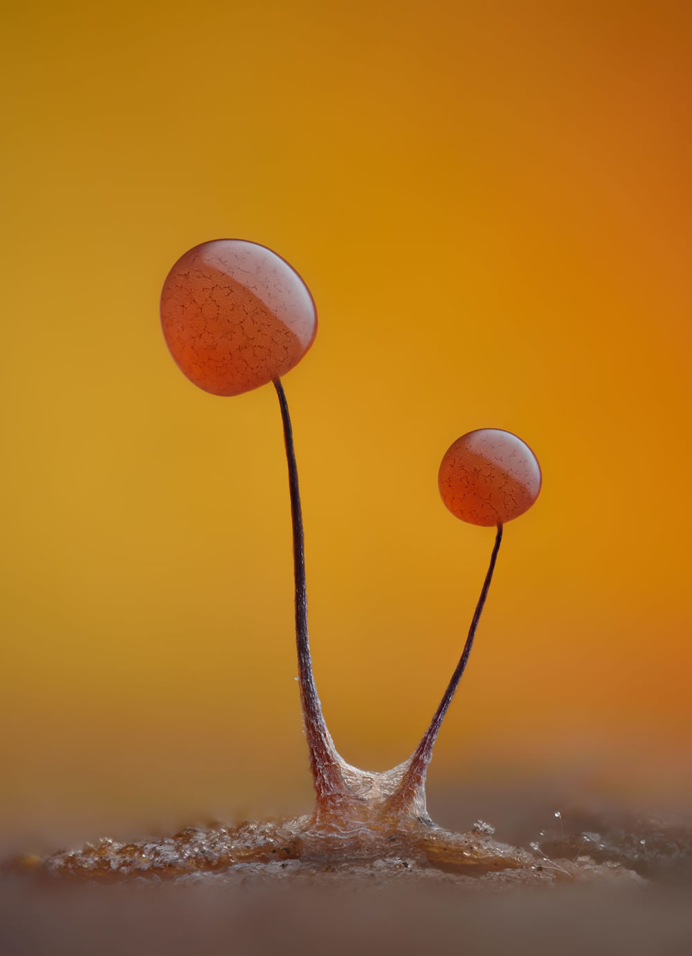
#6. Slime mold (Comatricha nigra) showing capillitial fibers through its translucent peridium by Timothy Boomer
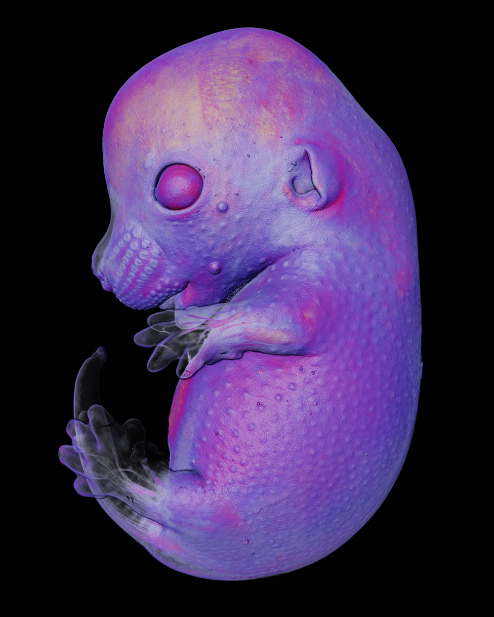
#7. Mouse embryo by Dr. Grigorii Timin & Dr. Michel Milinkovitch
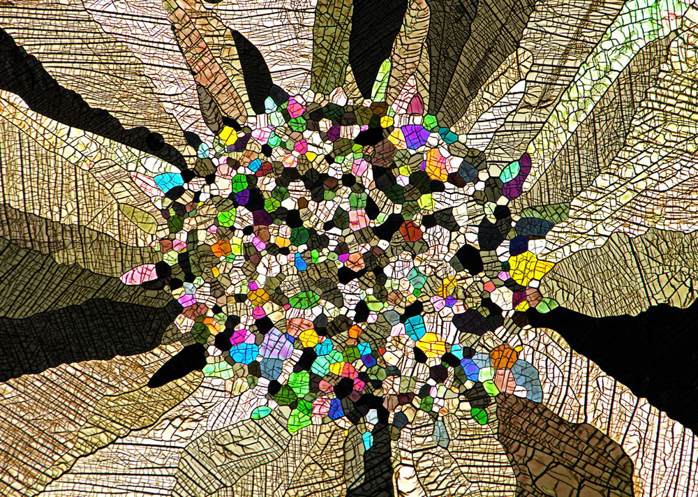
#8. Caffeine crystals by Stefan Eberhard
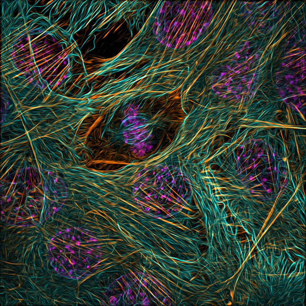
#9. Cytoskeleton of a dividing myoblast; tubulin (cyan), F-actin (orange) and nucleus (magenta) by Vaibhav Deshmukh
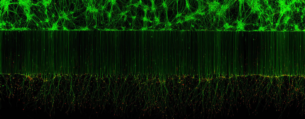
#10. Motor neurons grown in microfluidic device for separation of cell bodies (top) and axons (bottom). Green – microtubules; Red – growth cones (actin) by Melinda Beccari & Dr. Don W. Cleveland
#11. Crystallized sugar syrup by Dr. Diego García

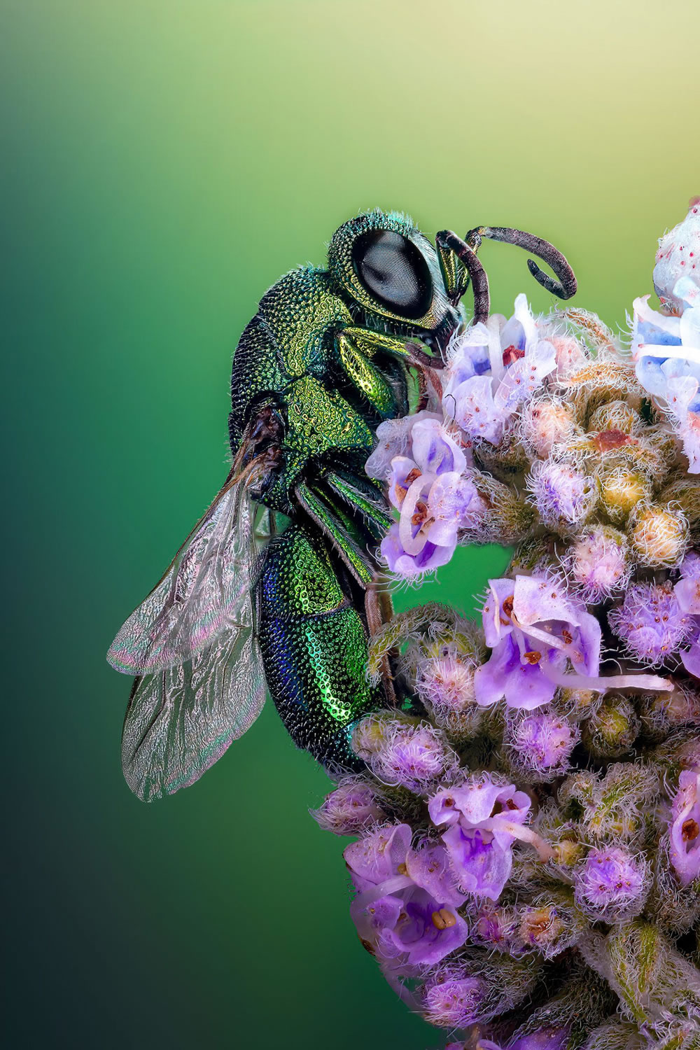
#12. Cuckoo wasp standing on a flower by Sherif Abdallah Ahmed
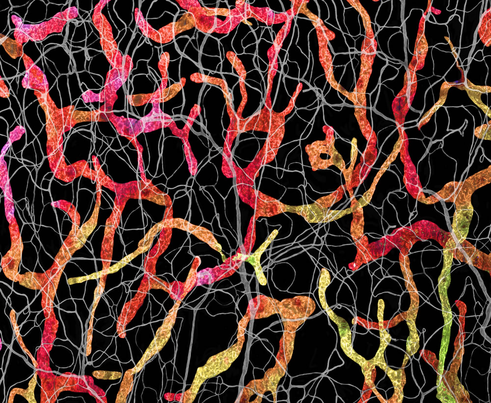
#13. Blood and lymphatic vasculatures in the ear skin of an adult mouse by Satu Paavonsalo & Dr. Sinem Karaman
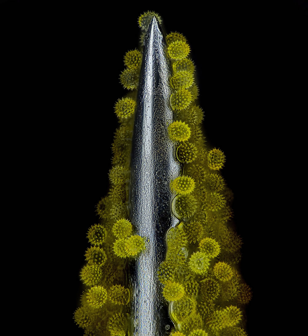
#14. Sunflower pollen on an acupuncture needle by John-Oliver Dum
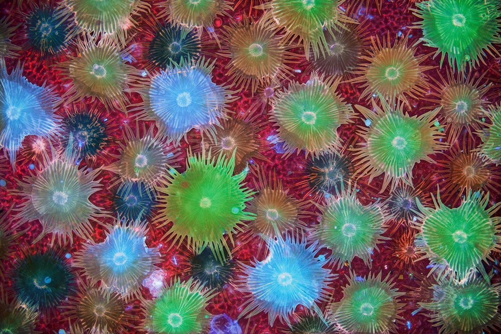
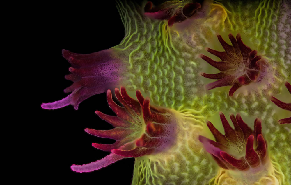
#15. Fluorescent image of an Acropora sp. showing individual polyps with symbiotic zooxanthellae by Dr. Pichaya Lertvilai
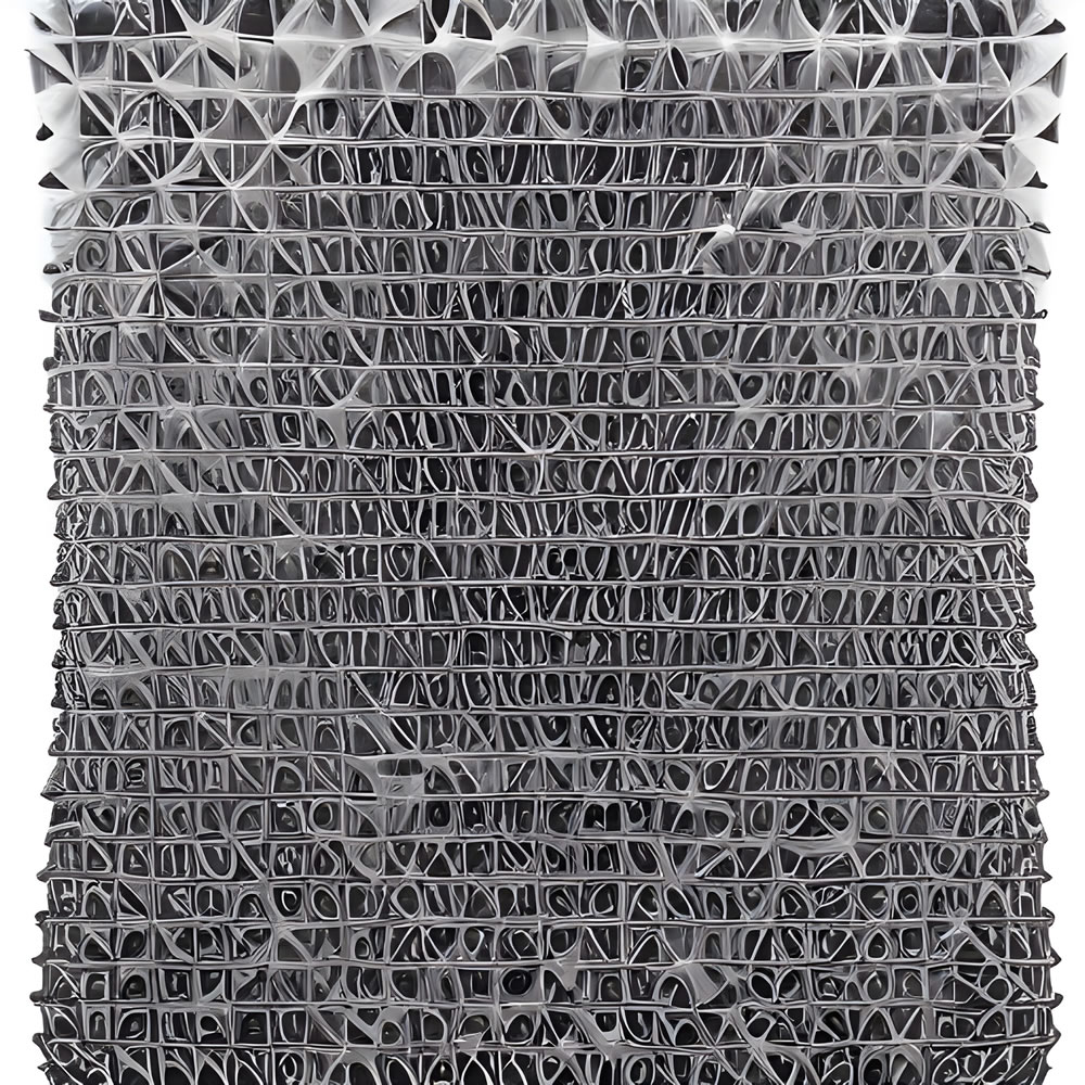
#16. Carbon nanotubes by Dr. Diego García
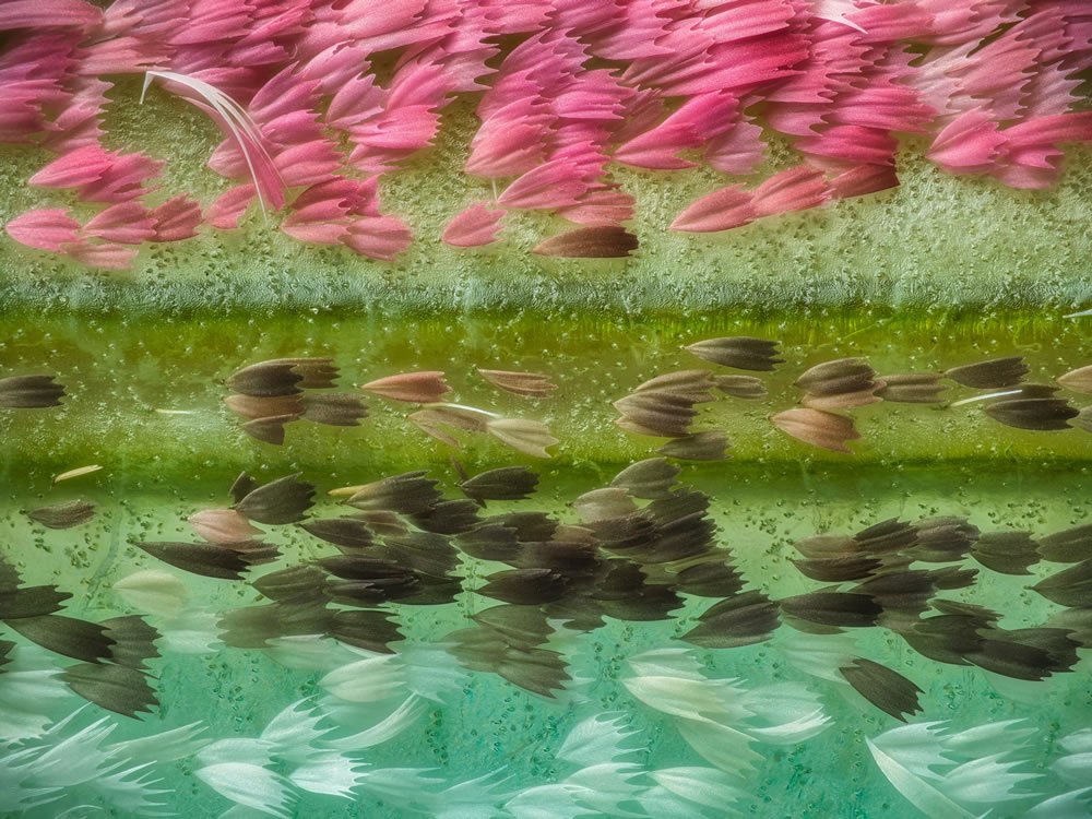
#17. Chinese moon moth (Actias ningpoana) wing scales by Yuan Ji
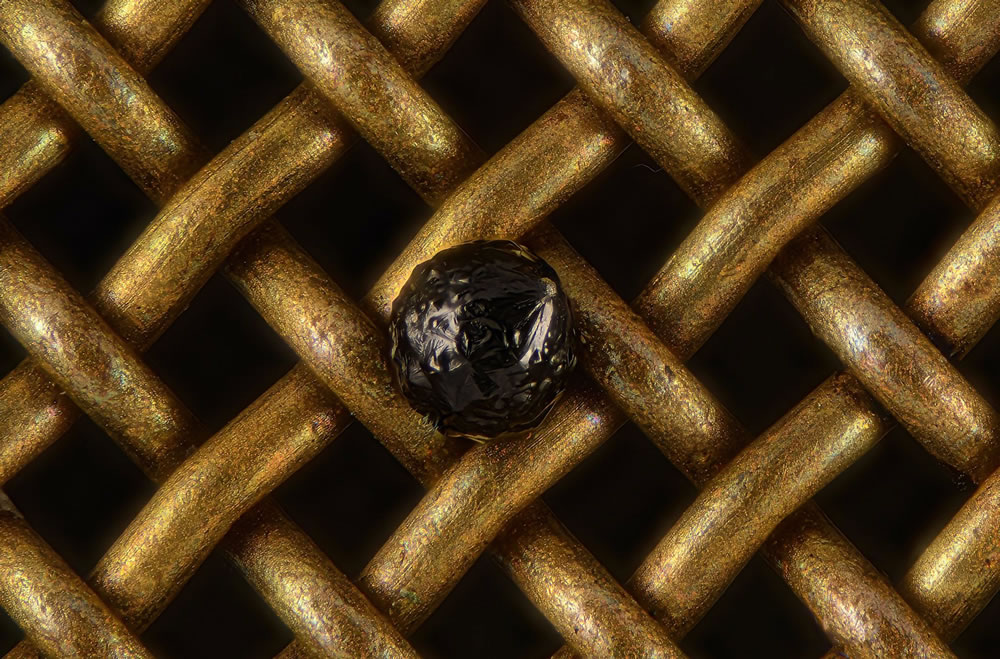
#18. A cryptocrystalline micrometeorite resting on a #80 testing sieve by Scott Peterson
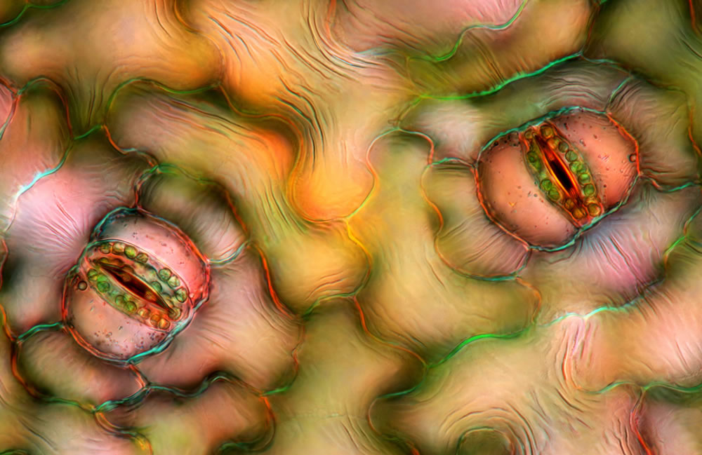
#19. Stomata in peace lily (Spathiphyllum sp.) leaf epidermis by Marek Miś
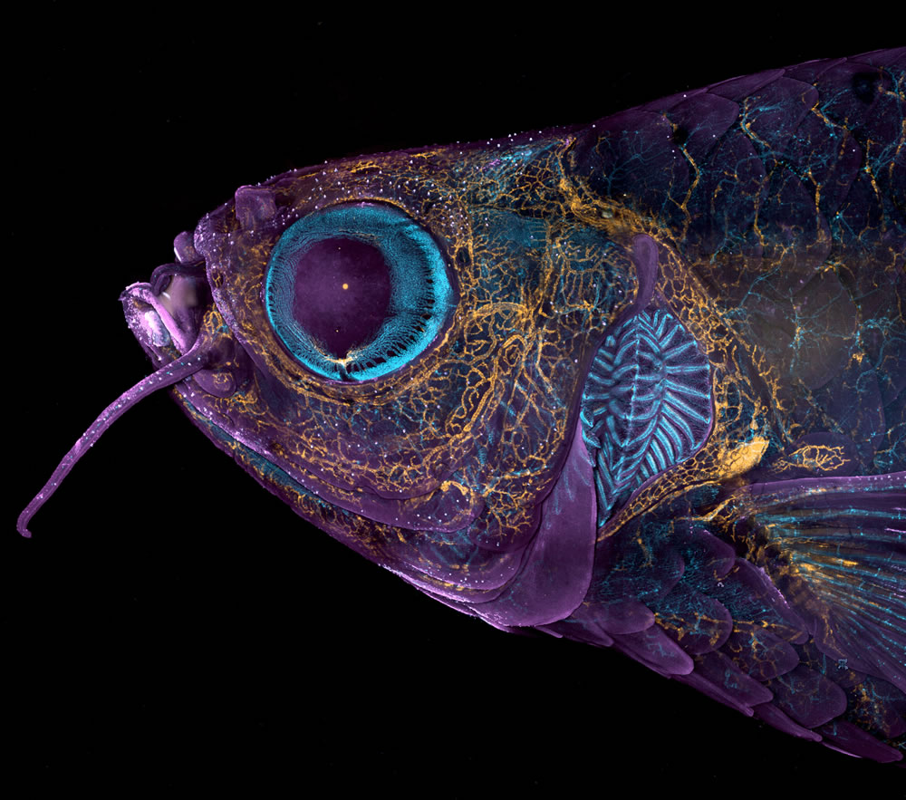
#20. Adult transgenic zebrafish head showing blood vessels (blue), lymphatic vessels (yellow), and the skin and scales (magenta) by Daniel Castranova & Dr. Brant M. Weinstein
