
Ever wondered what the world looks like when you zoom in way beyond what the naked eye can see? Get ready to have your mind blown! The Nikon Small World Photomicrography Competition is back with another incredible collection of images that turns the tiniest details into larger-than-life works of art.
For its 52nd annual competition, Nikon received 1,803 entries from photographers across 69 countries. From fascinating biological structures to everyday objects transformed into surreal masterpieces, this year’s winning photographs prove that beauty can hide in the most unexpected places.
Ole Bielfeldt, a student and experimental photographer based in Cologne, Germany, takes the top prize. His winning image captures an inkjet printer at a microscopic level, revealing an ordinary printing process in a completely unexpected way. Using a custom-built imaging rig, Bielfeldt transforms something we barely notice in daily life into a visually striking spectacle.
His creative project, macrofying, explores familiar objects through photomicroscopy, reminding us that even the most ordinary things can hold extraordinary secrets. These 42 award-winning photographs invite us to slow down, look closer, and discover a hidden universe that’s been right under our noses all along.
You can find more info about Nikon Small World:
Top 20 Photos
#1. 1st Place: Microscopic view of an inkjet printer depositing ink onto paper by Ole Bielfeldt
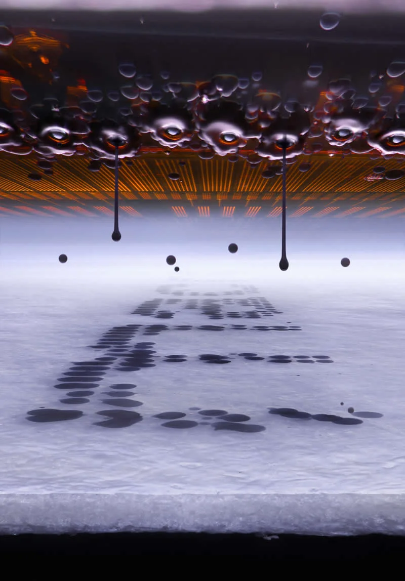
Technique: Slow-Motion, Image Stacking
Magnification: 20X (Objective Lens Magnification)
#2. 2nd Place: Jumping spider (Salticus scenicus) with pollen by John-Oliver Dum
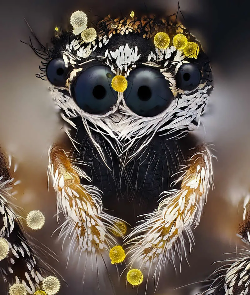
Technique: Image Stacking
Magnification: 20X (Objective Lens Magnification)
#3. 3rd Place: Neurons in an expanded mouse brain by Kaelin Wulf & Rachel Flannery
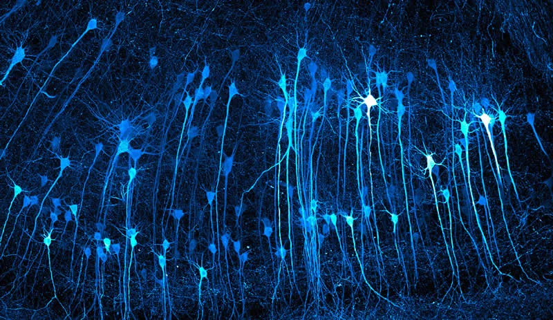
Technique: Expansion Microscopy, Fluorescence, Light Sheet
Magnification: 5X (Objective Lens Magnification)
#4. 4th Place: Tip of a ballpoint pen by Justin Theodore Benttinen
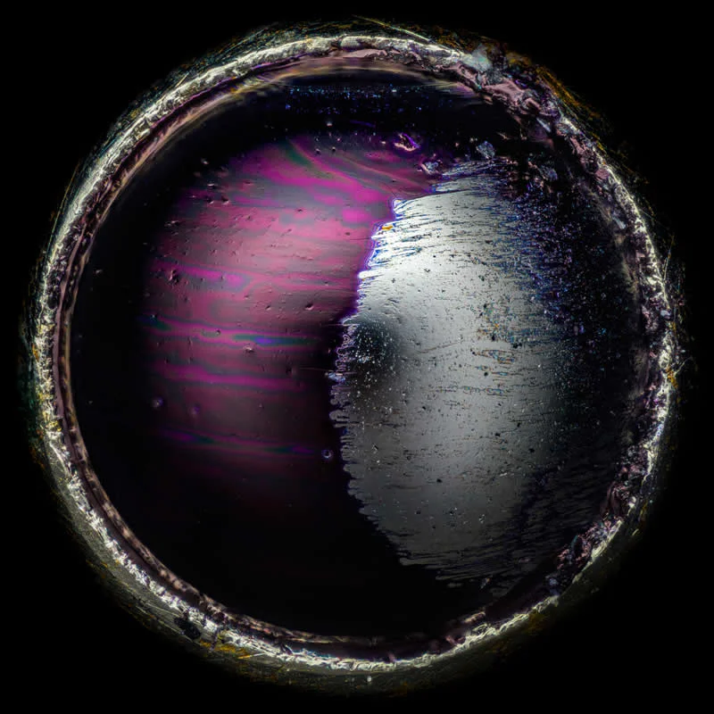
Technique: Image Stacking, Reflected Light
Magnification: 20X (Objective Lens Magnification)
#5. 5th Place: Inner structure of a closed daisy by Michael Crutchley
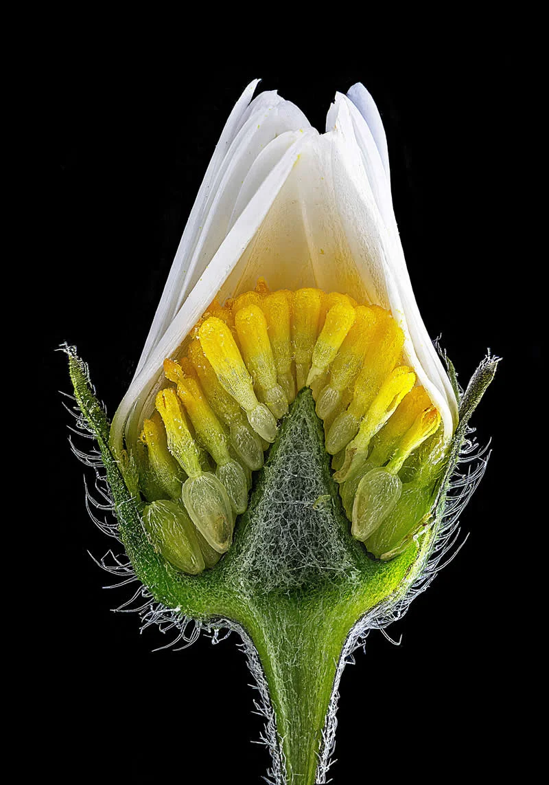
Location: Pembrokeshire, Southwest Wales, United Kingdom
Technique: Darkfield, Image Stacking
Magnification: 10X (Objective Lens Magnification)
#6. 6th Place: Hemitrichia slime mold spore structures (fruiting bodies) by Eric Epstein
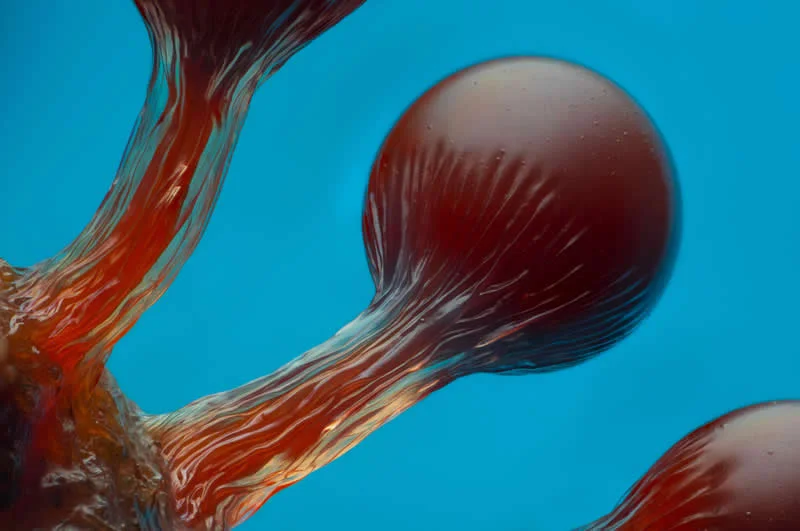
Location: Brooklyn, New York, USA
Technique: Image Stacking
Magnification: 10X (Objective Lens Magnification)
#7. 7th Place: Human neurons reprogrammed from skin cells growing in a microfluidic chamber by Dr. Bruno Cisterna &
Dr. Eric Vitriol
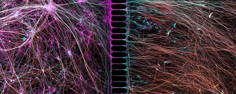
Technique: Confocal
Magnification: 20X (Objective Lens Magnification)
#8. 8th Place: Pencil lead during exposure to a high-voltage electrical discharge by Dr. Marcel Clemens
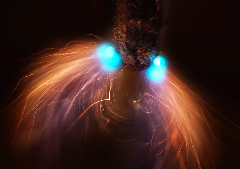
Location: Verona, Veneto, Italy
Technique: Image Stacking
Magnification: 10X (Objective Lens Magnification)
#9. 9th Place: Dedifferentiated liver endothelial cell by Dr. Francisco Lázaro-Diéguez
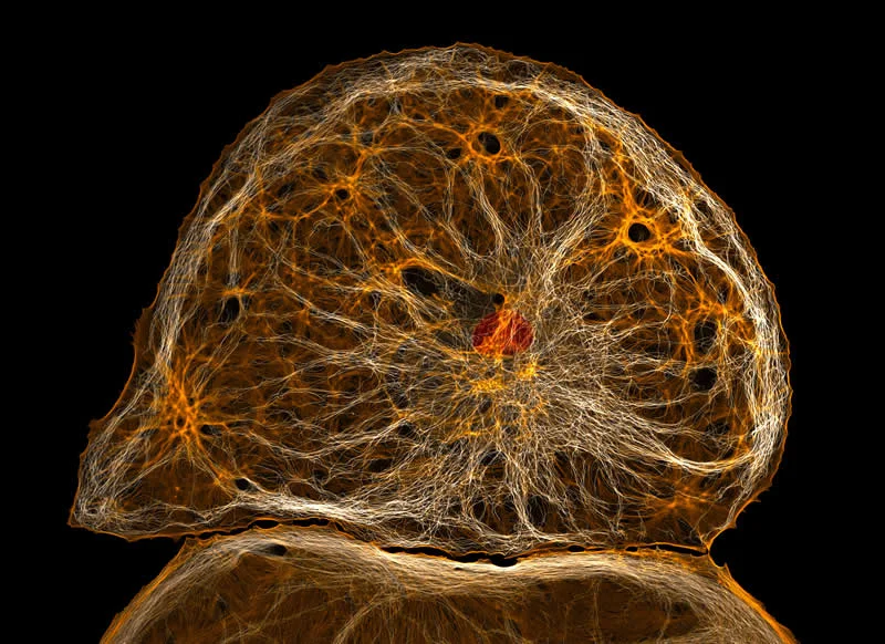
Technique: Confocal
Magnification: 40X (Objective Lens Magnification)
#10. 10th Place: A parasitoid wasp (Conura sp.) emerging from a butterfly chrysalis (Opsiphanes sp.) by Michael Parra Puentes
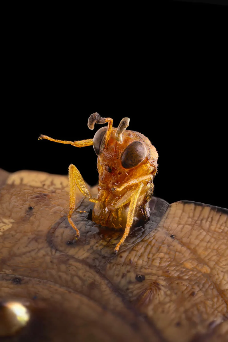
Technique: Image Stacking
Magnification: 3.7X (Objective Lens Magnification)
#11. 11th Place: 20 day old sea urchin larva with blue fluorescent nuclei by Svenja Carolin Kling
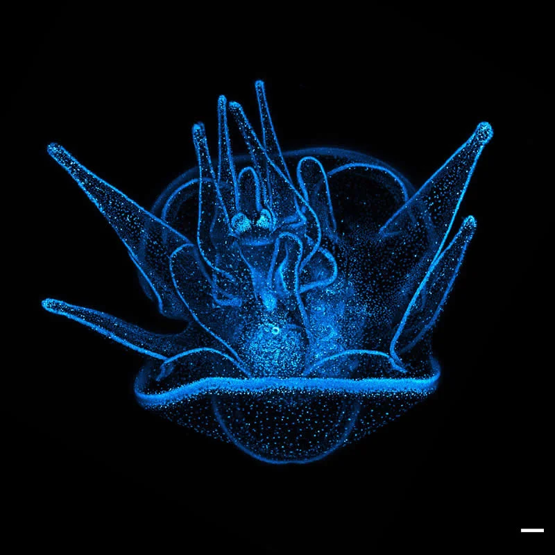
Technique: Confocal
Magnification: 20X (Objective Lens Magnification)
#12. 12th Place: A tardigrade molting and laying an egg in the old skin by Dr. Gonzalo Quiroga Artigas
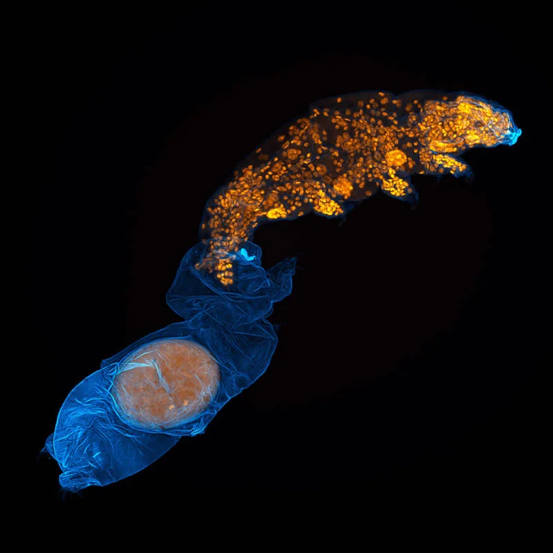
Technique: Confocal
Magnification: 40X (Objective Lens Magnification)
#13. 13th Place: Recrystallized estrogen by Sebastian Sparenga
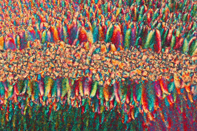
Technique: Polarized Light
Magnification: 10X (Objective Lens Magnification)
#14. 14th Place: Pilobolus fungi on sheep dung by Michael Crutchley
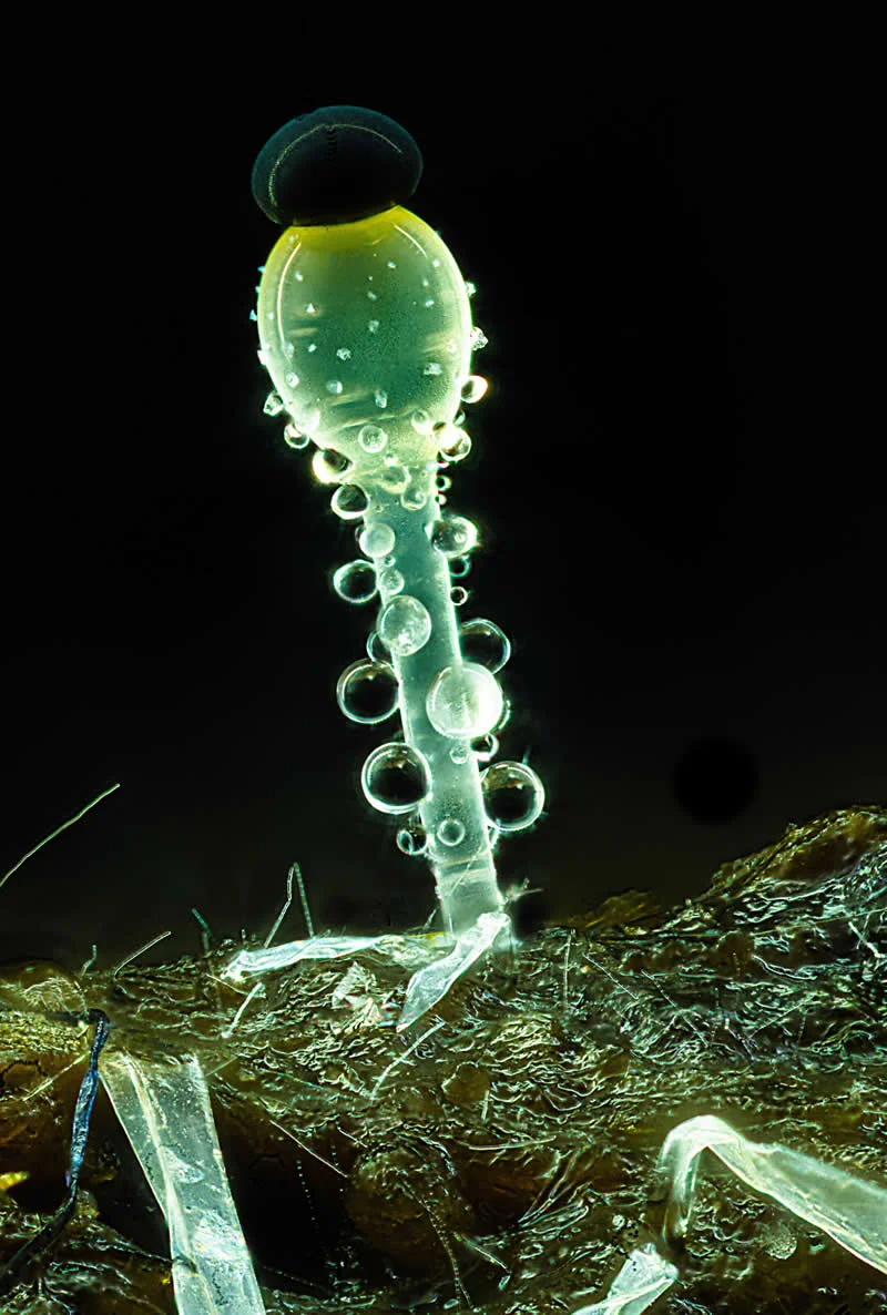
Location: Pembrokeshire, Southwest Wales, United Kingdom
Technique: Image Stacking
Magnification: 20X (Objective Lens Magnification)
#15. 15th Place: Mouse trachea showing tiny hair-like cells that move mucus and nearby nerves (blue) associated with the muscles (yellow) by Dr. Julien Resseguier
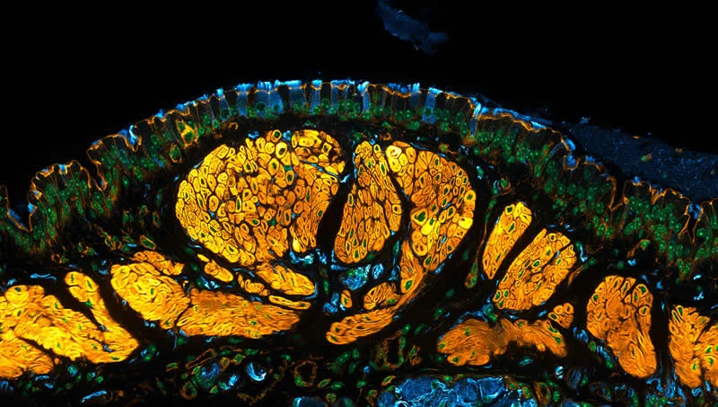
Technique: Confocal, Deconvolution, Fluorescence
Magnification: 60X (Objective Lens Magnification)
#16. 16th Place: Embryonic Xenopus skin cells, showing cilia (green), centrioles (red), and cell membranes (white) by
Pierre Nouaux & Laurent Kodjabachian, Camille Boutin
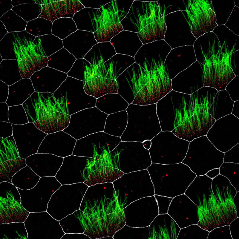
Technique: Confocal, Fluorescence
Magnification: 63X (Objective Lens Magnification)
#17. 17th Place: Lab-grown human intestinal tissue showing glowing, brick-like epithelial cell connections that form the intestine’s protective barrier by Dr. Gauri Kulkarni
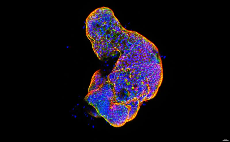
Technique: Confocal
Magnification: 20X (Objective Lens Magnification)
#18. 18th Place: 14-day-old mouse hippocampal neurons showing nuclei (green) and synaptic proteins (red) by Bilal Akhtar
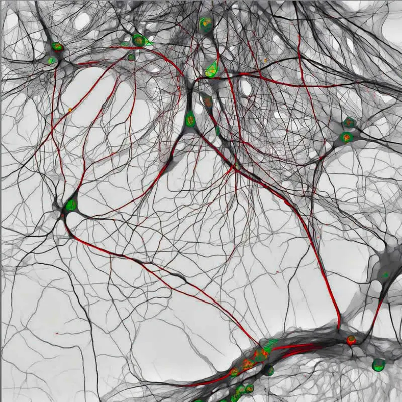
Technique: Confocal
Magnification: 40X (Objective Lens Magnification)
#19. 19th Place: Cross-section of a mouse eye showing retinal blood vessels and the optic nerve by Satu Paavonsalo &
Dr. Sinem Karaman
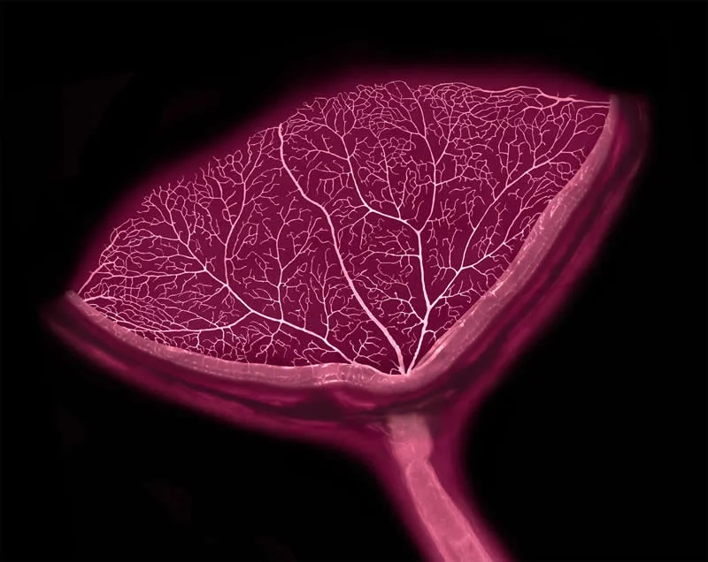
Technique: Confocal
Magnification: 20X (Objective Lens Magnification)
#20. 20th Place: Large sea snail (abalone) shell by Justin Theodore Benttinen
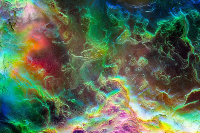
Technique: Image Stacking, Reflected Light
Magnification: 20X (Objective Lens Magnification)
Honorable Mentions
#21. Mouse (Mus musculus) uterus exhibiting epithelial gland structures by Claire J Ang
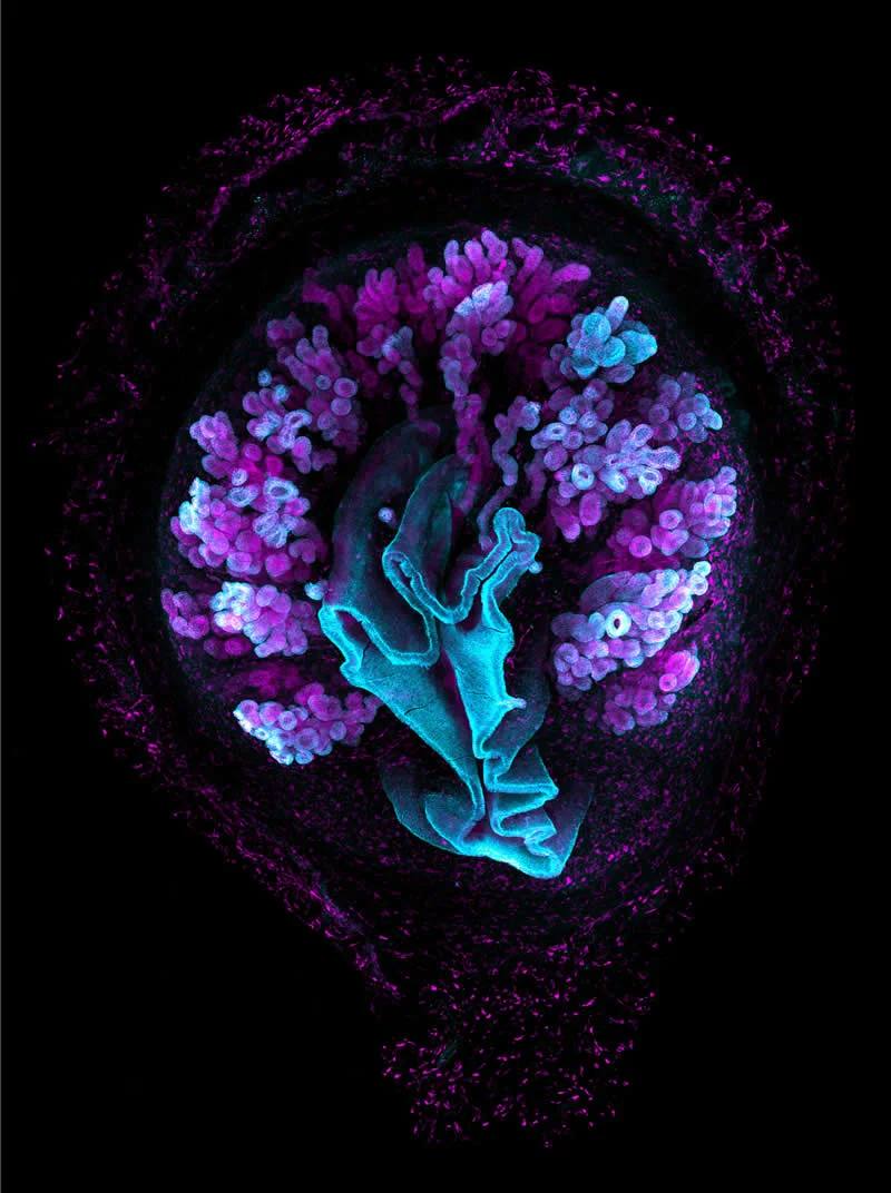
Technique: Confocal, Fluorescence, Image Stacking
Magnification: 10X (Objective Lens Magnification)
#22. A nest of intracellular parasites (Trypanosoma cruzi), growing inside human fibroblasts by Richard Atherton
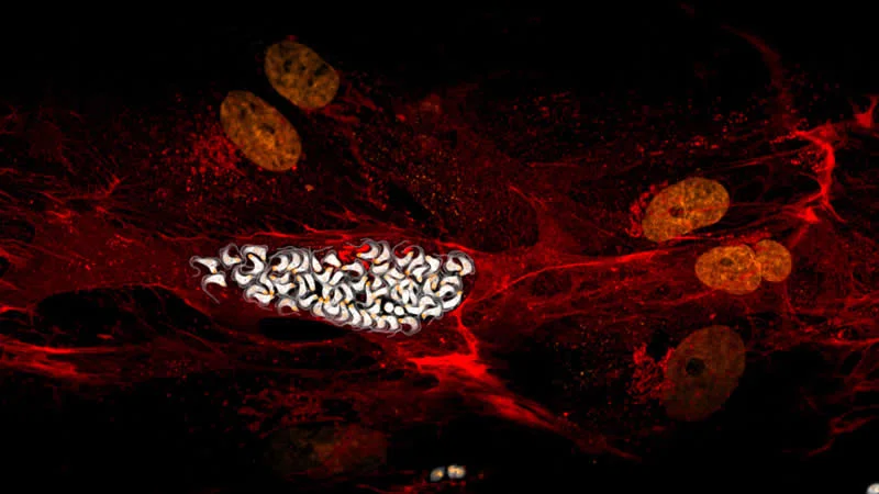
Technique: Confocal
Magnification: 100X (Objective Lens Magnification)
#23. African green monkey kidney cells (COS-7) displaying the microtubule network (cyan) and mitochondria (red and yellow) by Walid Idi
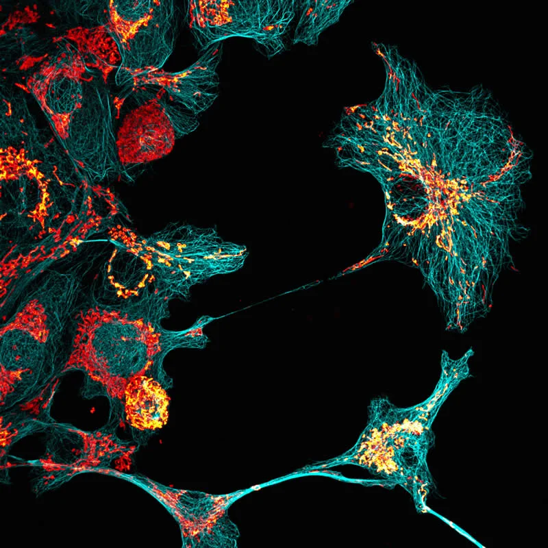
Technique: Confocal
Magnification: 40X (Objective Lens Magnification)
#24. A common daisy flower head (Bellis perennis) glowing with its natural autofluorescence by Jan Martinek
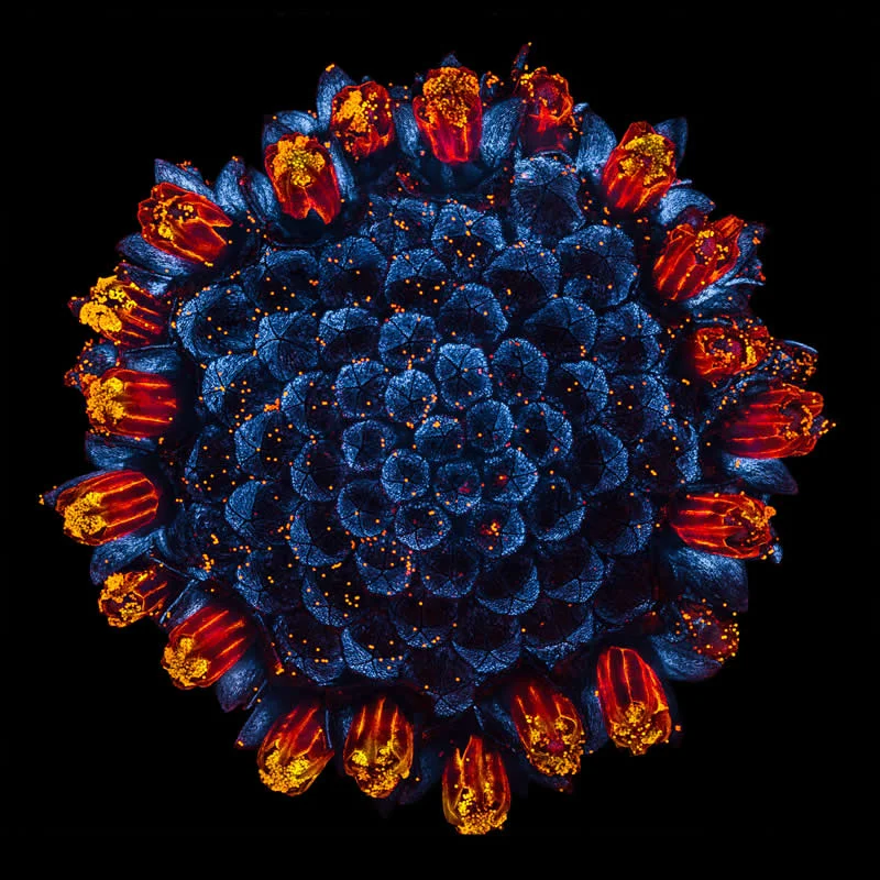
Technique: Confocal
Magnification: 4X (Objective Lens Magnification)
#25. An Arabidopsis root coil by Jan Martinek
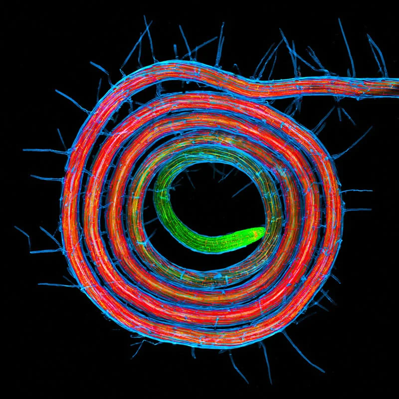
Technique: Confocal
Magnification: 5X (Objective Lens Magnification)
#26. Neurons of red-eared slider (Trachemys scripta) embryo by Amartya Tashi Mitra, Paul Maier, Aria Huang, Nathaniel Sweet, Anthony Wokasch
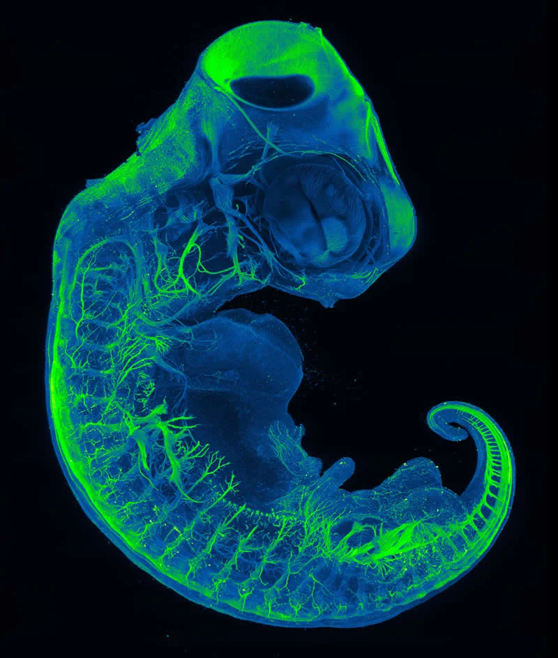
Technique: Light Sheet
Magnification: 9X (Objective Lens Magnification)
#27. Seal louse (Antarctophthirus sp.) gripping a single seal hair by Brandon Antonio Segura Torres, Priscilla Vieto Bonilla, Paula Alejandra Olivera
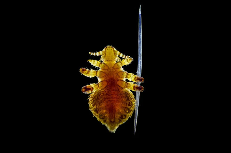
Technique: Darkfield, Image Stacking
Magnification: 100X (Objective Lens Magnification)
#28. Live plant roots (Arabidopsis thaliana, green) colonized by bacteria (Bacillus thuringiensis, magenta) by Dr. Kevin Y Shih
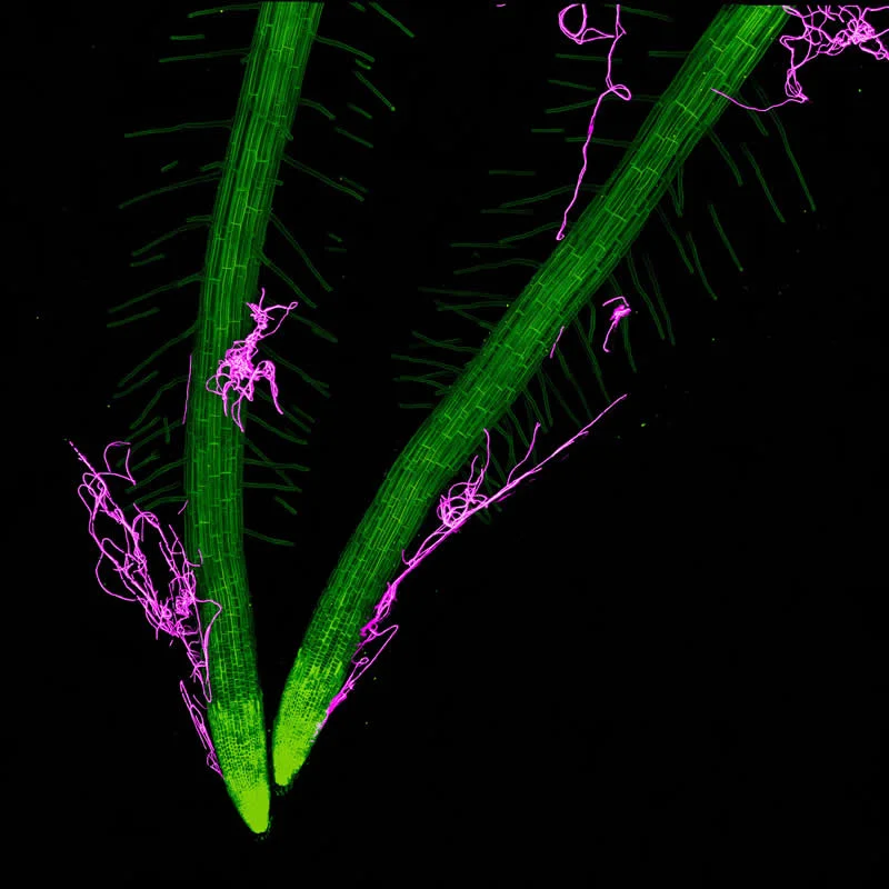
Technique: Confocal
Magnification: 40X (Objective Lens Magnification)
#29. A pair of elongated Stentor coeruleus cells during mating. The cells are fused at anterior edge of their mouth to allow for the transfer of DNA. by Dr. Mark Slabodnick
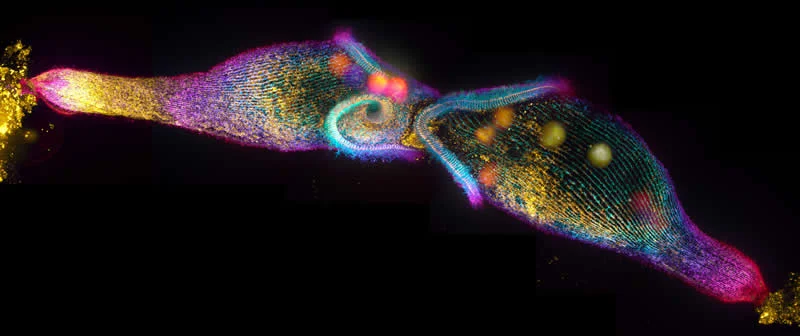
Technique: Confocal, Fluorescence, Image Stacking, Image Stitching, Depth-coloration
Magnification: 63X (Objective Lens Magnification)
Image of Distinction
#30. Cytoskeleton stain of the “jumping” ciliate Halteria grandinella, a common pondwater protist by Rocky An,
Dr. Manu Prakash
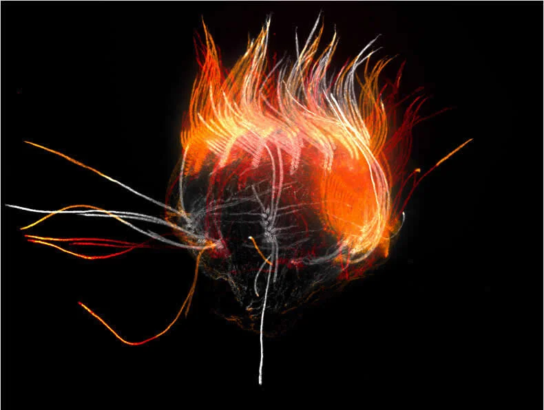
Technique: Confocal, Fluorescence, Expansion Microscopy
Magnification: 100X (Objective Lens Magnification)
#31. Nerves and vasculature connecting the ovary, fallopian tubes, and uterus in a mouse (Mus musculus) by Claire J Ang
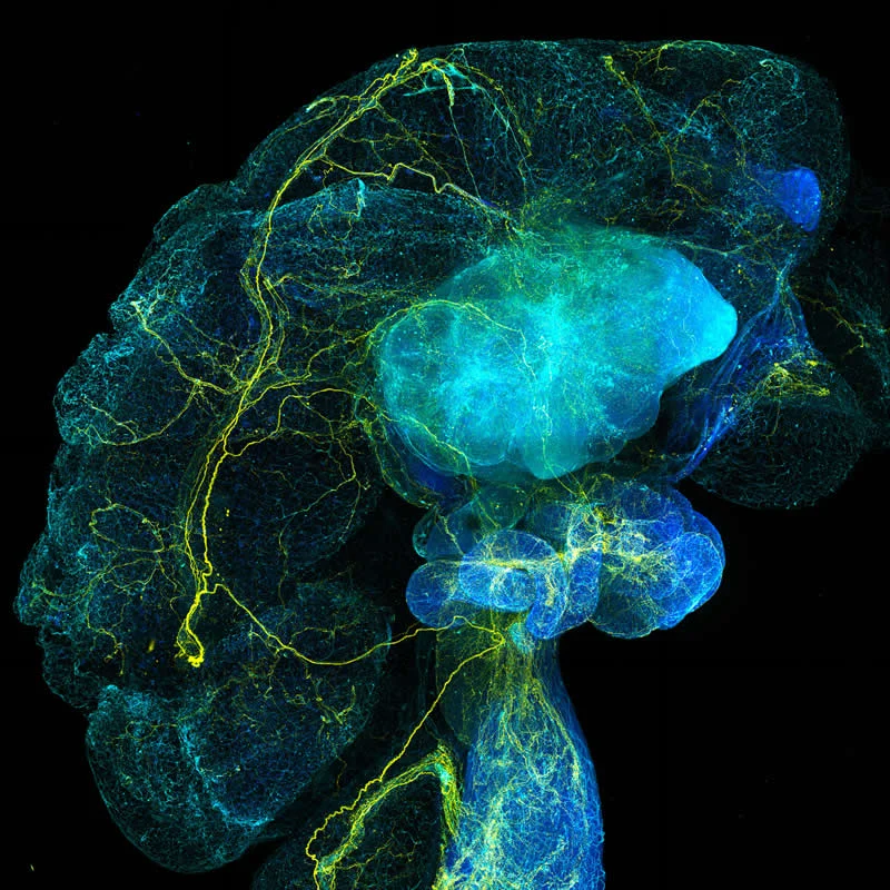
Technique: Confocal, Fluorescence, Image Stacking
Magnification: 10X (Objective Lens Magnification)
#32. Naturally water etched spessartine garnet from Brazil by Justin Theodore Benttinen
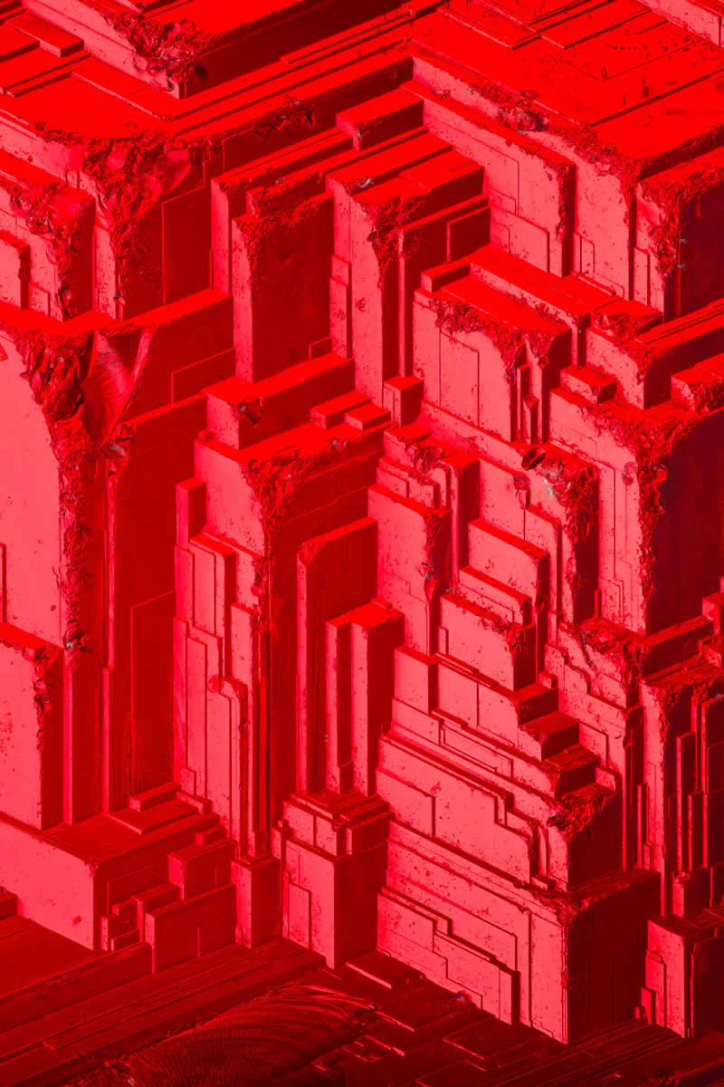
Technique: Transmitted Light, Image Stacking, Reflected Light
Magnification: 20X (Objective Lens Magnification)
#33. Paired zebrafish embryos by Dr. Dylan T. Burnette & Dr. James Hayes
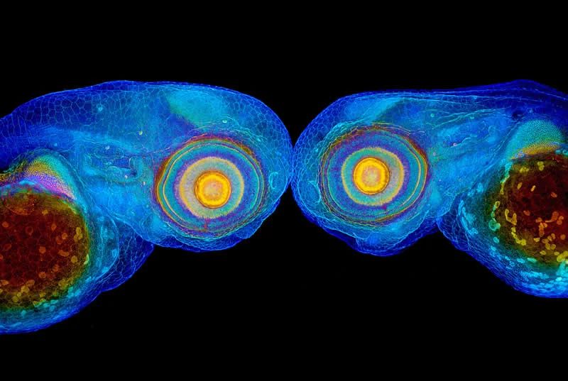
Technique: Structured Illumination Microscopy (SIM)
Magnification: 60X (Objective Lens Magnification)
#34. Intestinal tuft cell by Dr. Amy Engevik
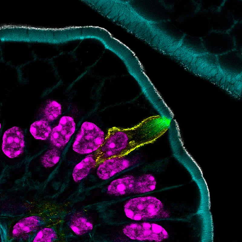
Technique: Confocal
Magnification: 63X (Objective Lens Magnification)
#35. Human neurons (differentiated from induced pluripotent stem cells) by Faith Fernandes, Dr. James Glover
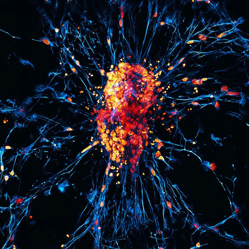
Technique: Confocal
Magnification: 60X (Objective Lens Magnification)
#36. Hibiscus flower with pollen by Frank Fox
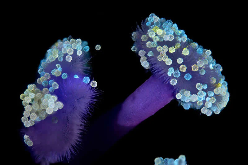
Technique: Fluorescence
Magnification: 10X (Objective Lens Magnification)
#37. Western honey bee (Apis mellifera) with pollen by Giuseppe Franchina
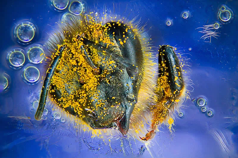
Location: Messina, Italy
Technique: Image Stacking
Magnification: 4X (Objective Lens Magnification)
#38. Neurons connecting to sensory cells of the inner ear in culture by Dr. Stephen Freeman
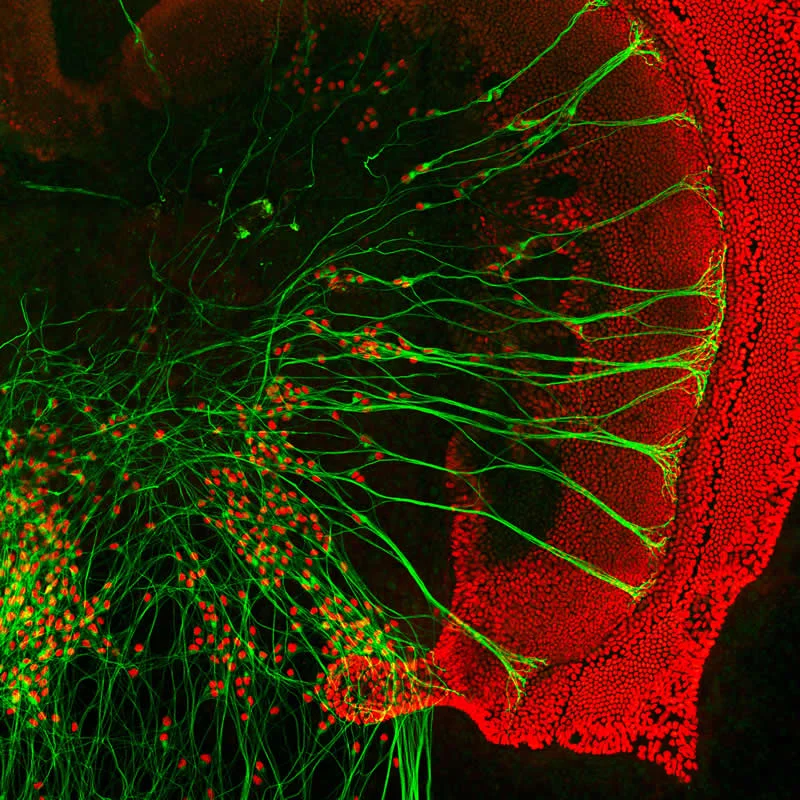
Technique: Confocal
Magnification: 20X (Objective Lens Magnification)
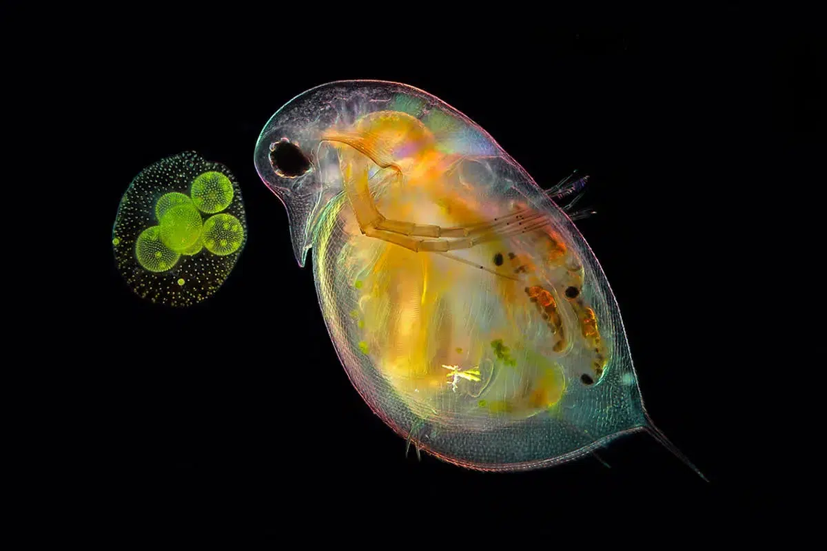
#39. Daphnia sp. (water flea) and Volvox sp. (green algae) by Anne Gleich
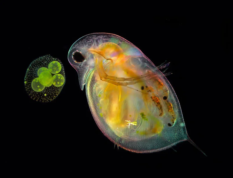
Technique: Darkfield, Image Stacking, Polarized Light
Magnification: 4X (Objective Lens Magnification)
#40. A kidney stone showing two formations: a smoother mineral buildup and sharp crystalline growth by Saulius Gugis
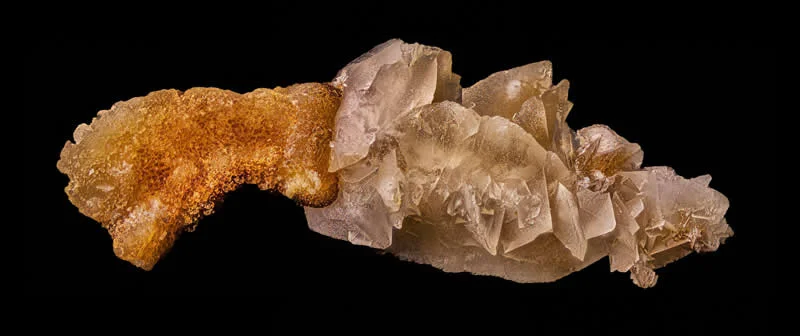
Location: Naperville, Illinois, USA
Technique: Image Stacking
Magnification: 10X (Objective Lens Magnification)
#41. Developing pupil and optic nerve tract of a Zebrafish embryo by Dr. James Hayes
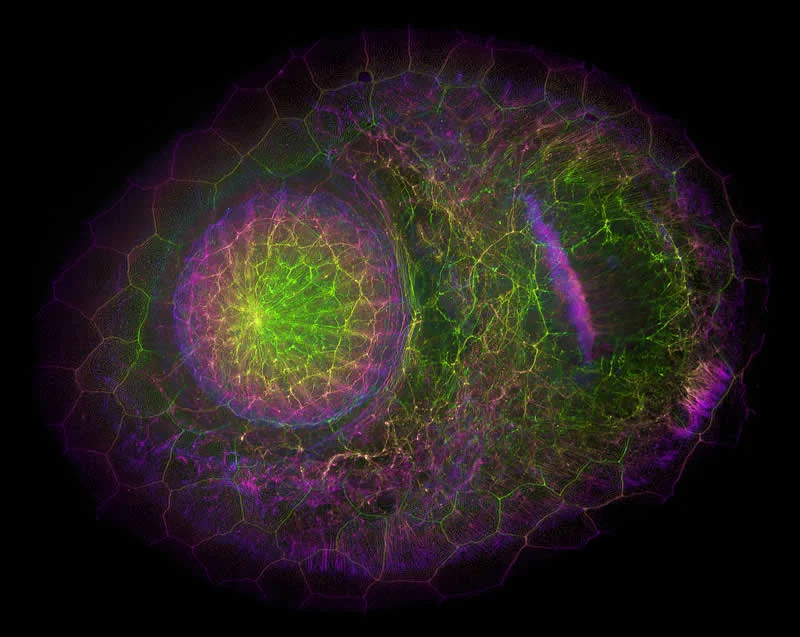
Technique: Confocal
Magnification: 60X (Objective Lens Magnification)
#42. Simultaneous cell division in a 2-cell mouse embryo by Blake Hernandez
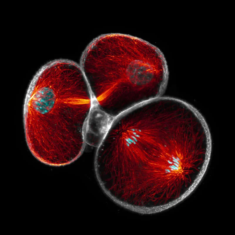
Technique: Confocal
Magnification: 40X (Objective Lens Magnification)
Frequently Asked Questions (FAQ):
What is the Nikon Small World Photomicrography Competition?
The Nikon Small World Photomicrography Competition is an annual international contest celebrating extraordinary images captured through microscopes. It brings together photographers and scientists who reveal fascinating details of nature, biological structures, and everyday objects invisible to the naked eye.
How many entries were submitted to Nikon Small World?
The 52nd annual Nikon Small World Photomicrography Competition received 1,803 entries from photographers representing 69 countries. The impressive international participation showcases the growing appeal of photomicrography and the remarkable creativity involved in capturing microscopic subjects.
Who won the Nikon Small World grand prize?
Ole Bielfeldt, a student and experimental photographer based in Cologne, Germany, won first place. His image shows an inkjet printer at a microscopic level, turning an ordinary printing process into an extraordinary visual experience with specialized imaging equipment.
What is photomicrography?
Photomicrography is the art and science of photographing subjects through a microscope. It reveals intricate structures, textures, and patterns that are usually invisible to the human eye, making it possible to explore everything from tiny organisms to unexpected details in everyday materials.
How did Ole Bielfeldt capture his winning photograph?
Ole Bielfeldt used a custom-built imaging rig to photograph an inkjet printing process at a microscopic scale. Through his creative project, macrofying, he explores familiar objects from unusual perspectives, demonstrating how specialized imaging techniques can transform ordinary subjects into captivating photographs.